Select From The Following for More Details
Back to Topics<<<<

Tyrosine-Derived Neurotransmitters
The majority of tyrosine that does not get incorporated into proteins is catabolized for energy production. One other significant fate of tyrosine is conversion to the catecholamines. The catecholamine neurotransmitters are dopamine, norepinephrine, and epinephrine (see also Biochemistry of Nerve Transmission).
Norepinephrine is the principal neurotransmitter of sympathetic postganglionic endings. Both norepinephrine and the methylated derivative, epinephrine are stored in synaptic knobs of neurons that secrete it, however, epinephrine is not a mediator at postganglionic sympathetic endings.
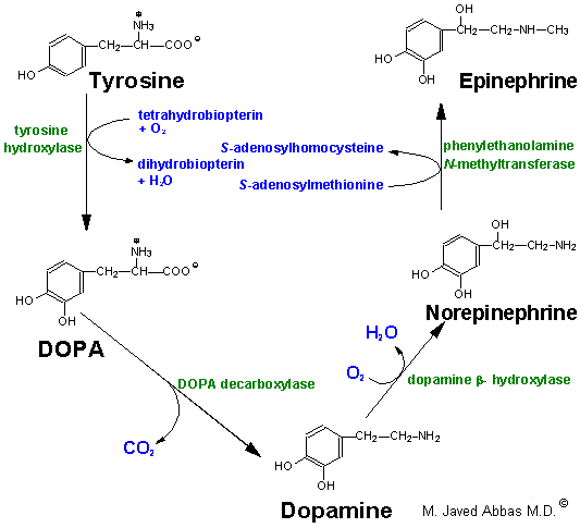
Tyrosine is transported into catecholamine-secreting neurons and adrenal medullary cells where catechaolamine synthesis takes place. The first step in the process requires tyrosine hydroxylase, which like phenylalanine hydroxylase requires tetrahydrobiopterin as cofactor. The hydroxylation reaction generates DOPA (3,4-dihydrophenylalanine). DOPA decarboxylase converts DOPA to dopamine, dopamine b-hydroxylase converts dopamine to norepinephrine and phenylethanolamine N-methyltransferase converts norepinephrine to epinephrine. This latter reaction is one of several in the body that uses SAM as a methyl donor generating S-adenosylhomocysteine. Within the substantia nigra and some other regions of the brain, synthesis proceeds only to dopamine. Within the adrenal medulla dopamine is converted to norepinephrine and epinephrine.

Synthesis of the catecholamines from tyrosine.

Once synthesized, dopamine, norepinephrine and epinephrine are packaged in granulated vesicles. Within these vesicles, norepinephrine and epinephrine are bound to ATP and a protein called chromogranin A.
Metabolism of the catecholemines occurs through the actions of catecholamine-O-methyltransferase, (COMT) and monoamine oxidase, (MAO). Both of these enzymes are widley distributed throughout the body. However, COMT is not found in nerve endings as is MAO.
back to the top
Tryptophan-Derived Neurotransmitters
Tryptopan serves as the precursor for the synthesis of serotonin (5-hydroxytryptamine, 5-HT, see also Biochemistry of Nerve Transmission) and melatonin (N-acetyl-5-methoxytryptamine).

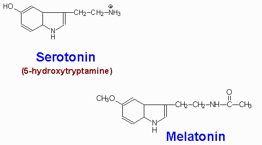
Serotonin is synthesized through 2-step process involving a tetrahydrobiopterin-dependent hydroxylation reaction (catalyzed by tryptophan-5-monooxygenase) and then a decarboxylation catalyzed by aromatic L-amino acid decarboxylase. The hydroxylase is normally not saturated and as a result, an increased uptake of tryptophan in the diet will lead to increased brain serotonin content.
Serotonin is present at highest concentrations in platelets and in the gastrointestinal tract. Lesser amounts are found in the brain and the retina. Serotonin containing neurons have their cell bodies in the midline raphe nuclei of the brain stem and project to portions of the hypothalamus, the limbic system, the neocortex and the spinal cord. After release from serotonergic neurons, most of the released serotonin is recaptured by an active reuptake mechanism. The function of the antidepressant, Prozac is to inhibit this reuptake process, thereby, resulting in prolonged serotonin presence in the synaptic cleft.
The function of serotonin is exerted upon its interaction with specific receptors. Several serotonin receptors have been cloned and are identified as 5HT1, 5HT2, 5HT3, 5HT4, 5HT5, 5HT6, and 5HT7. Within the 5HT1 group there are subtypes 5HT1A, 5HT1B, 5HT1D, 5HT1E, and 5HT1F. There are three 5HT2 subtypes, 5HT2A, 5HT2B, and 5HT2C as well as two 5HT5 subtypes, 5HT5a and 5HT5B. Most of these receptors are coupled to G-proteins that affect the activities of either adenylate cyclase or phospholipase Cg (PLCg). The 5HT3 class of receptors are ion channels.
Some serotonin receptors are presynaptic and others postsynaptic. The 5HT2A receptors mediate platelet aggregation and smooth muscle contraction. The 5HT2C receptors are suspected in control of food intake as mice lacking this gene become obese from increased food intake and are also subject to fatal seizures. The 5HT3 receptors are present in the gastrointestinal tract and are related to vomiting. Also present in the gastrointestinal tract are 5HT4 receptors where they function in secretion and peristalsis. The 5HT6 and 5HT7 receptors are distributed throughout the limbic system of the brain and the 5HT6 receptors have high affinity for antidepressant drugs.
Melatonin is derived from serotonin within the pineal gland and the retina, where the necessary N-acetyltransferase enzyme is found. The pineal parenchymal cells secrete melatonin into the blood and cerebrospinal fluid. Synthesis and secretion of melatonin increases during the dark period of the day and is maintained at a low level during daylight hours. This diurnal variation in melatonin synthesis is brought about by norepinephrine secreted by the postganglionic sympathetic nerves that innervate the pineal gland. The effects of norepinephrine are exerted through interaction with b-adrenergic receptors. This leads to increased levels of cAMP, which in turn activate the N-acetyltransferase required for melatonin synthesis. Melatonin functions by inhibiting the synthesis and secretion of other neurotransmitters such as dopamine and GABA.
back to the top
Creatine Biosynthesis
Creatine is synthesized in the liver by methylation of guanidoacetate using SAM as the methyl donor. Guanidoacetate itself is formed in the kidney from the amino acids arginine and glycine.

Synthesis of creatine and creatinine

Creatine is used as a storage form of high energy phosphate. The phosphate of ATP is transferred to creatine, generating creatine phosphate, through the action of creatine phosphokinase. The reaction is reversible such that when energy demand is high (e.g. during muscle exertion) creatine phosphate donates its phosphate to ADP to yield ATP.
Both creatine and creatine phosphate are found in muscle, brain and blood. Creatinine is formed in muscle from creatine phosphate by a nonenzymatic dehydration and loss of phosphate. The amount of creatinine produced is related to muscle mass and remains remarkably constant from day to day. Creatinine is excreted by the kidneys and the level of excretion (creatinine clearance rate) is a measure of renal function.
back to the top
Glutathione Functions
Glutathione (abbreviated GSH) is a tripeptide composed of glutamate, cysteine and glycine that has numerous important functions within cells. It serves as a reductant, is conjugated to drugs to make them more water soluble, is involved in amino acid transport across cell membranes (the g-glutamyl cycle), is a part of the peptidoleukotrienes, serves as a cofactor for some enzymatic reactions and as an aid in the rearrangement of protein disulfide bonds.

Synthesis of glutathione (GSH)Structure of GSSG

The role of GSH as a reductant is extremely important particularly in the highly oxidizing environment of the erythrocyte. The sulfhydryl of GSH can be used to reduce peroxides formed during oxygen transport. The resulting oxidized form of GSH consists of two molecules disulfide bonded together (abbreviated GSSG). The enzyme glutathione reductase utilizes NADPH as a cofactor to reduce GSSG back to two moles of GSH. Hence, the pentose phosphate pathway is an extremely important pathway of erythrocytes for the continuing production of the NADPH needed by glutathione reductase. In fact as much as 10% of glucose consumption, by erythrocytes, may be mediated by the pentose phosphate pathway.
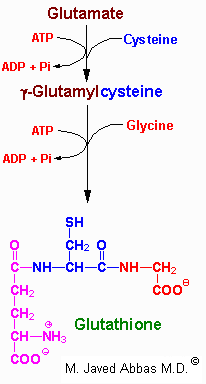
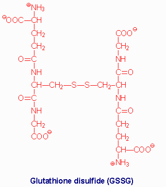
Several mechanisms exist for the transport of amino acids across cell membranes. Many are symport or antiport mechanisms that couple amino acid transport to sodium transport. The g-glutamyl cycle is an example of a group transfer mechanism of amino acid transport. Although this mechanism requires more energy input, it is rapid and has a high capacity. The cycle functions primarily in the kidney, particularly renal epithelial cells. The enzyme g-glutamyl transpeptidase is located in the cell membrane and shuttles GSH to the cell surface to interact with an amino acid. Reaction with an amino acid liberates cysteinylglycine and generates a g-glutamyl-amino acid which is transported into the cell and hydrolyzed to release the amino acid. Glutamate is released as 5-oxoproline and the cysteinylglycine is cleaved to its component amino acids. Regeneration of GSH requires an ATP-dependent conversion of 5-oxoproline to glutamate and then the 2 additional moles of ATP that are required during the normal generation of GSH.
back to the top
Polyamnine Biosynthesis
One of the earliest signals that cells have entered their replication cycle is the appearance of elevated levels of mRNA for ornithine decarboxylase (ODC), and then increased levels of the enzyme, which is the first enzyme in the pathway to synthesis of the polyamines. Because of the latter, and because the polyamines are highly cationic and tend to bind nucleic acids with high affinity, it is believed that the polyamines are important participants in DNA synthesis, or in the regulation of that process.

The key features of the pathway are that it involves putrescine, an ornithine catabolite, and S-adenosylmethionine (SAM) as a donor of 2 propylamine residues. The first propylamine conjugation yields spermidine and addition of another to spermidine yields spermine.
The function of ODC is to produce the 4-carbon saturated diamine, putrescine. At the same time, SAM decarboxylase cleaves the SAM carboxyl residue, producing decarboxylated SAM (S-adenosymethylthiopropylamine), which retains the methyl group usually involved in SAM methyltransferase activity. SAM decarboxylase activity is regulated by product inhibition and allosterically stimulated by putrescine. Spermidine synthase catalyzes the condensation reaction, producing spermidine and 5'-methylthioadenosine. A second propylamine residue is added to spermidine producing spermine.
The signal for regulating ODC activity is unknown, but since the product of its activity, putrescine, regulates SAM decarboxylase activity, it appears that polyamine production is principally regulated by ODC concentration.
The butylamino group of spermidine is used in a posttranslational modification reaction important to the process of translation. A specific lysine residue in the translational initiation factor eIF-4D is modified. Following the modification the residue is hydroxylated yielding a residue in the protein termed hypusine.
back to the top
Nitric Oxide Synthesis and Function
Vasodilators, such as acetylcholine, do not exert their effects upon the vascular smooth muscle cell in the absence of the overlying endothelium. When acetylcholine binds its receptor on the surface of endothelial cells, a signal cascade, coupled to the activation phospholipase C-g (PLCg), is initiated. The PLCg-mediated release of inositol trisphosphate, IP3 (from membrane associated phosphatidylinositol-4,5-bisphosphate, PIP2), leads to the release of intracellular stores of Ca2+. In turn, the elevation in Ca2+ leads to the liberation of endothelium-derived relaxing factor (EDRF) which then diffuses into the adjacent smooth muscle. Within smooth muscle cells, EDRF reacts with the heme moiety of a soluble guanylyl cyclase, resulting in activation of the latter and a consequent elevation of intracellular levels of cGMP. The net effect is the activation of cGMP-responsive enzymes which lead to smooth muscle cell relaxation. The coronary artery vasodilator, nitroglycerin, acts to increase intracellular release of EDRF and thus of cGMP.
Quite unexpectedly, EDRF was found to be the free radical diatomic gas, nitric oxide, NO. NO is formed by the action of NO synthase, (NOS) on the amino acid arginine.

arginine ----->  citrulline + NO

Nitric oxide is involved in a number of other important cellular processes in addition to its impact on vascular smooth muscle cells. Events initiated by NO that are important for blood coagulation include inhibition of platelet aggregation and adhesion and inhibition of neutrophil adhesion to platelets and to the vascular endothelium. NO is also generated by cells of the immune system and as such is involved in non-specific host defense mechanisms and macrophage-mediated killing. NO also inhibits the proliferation of tumor cells and microorganisms. Additional cellular responses to NO include induction of apoptosis (programmed cell death), DNA breakage and mutation.
NOS is a very complex enzyme, employing five redox cofactors: NADPH, FAD, FMN, heme and tetrahydrobiopterin. NO can also be formed from nitrite, derived from vasodilators such as glycerin trinitrate during their metabolism. The half-life of NO is extremely short, lasting only 2-4 seconds. This is because it is a highly reactive free radical and interacts with oxygen and superoxide. NO is inhibited by hemoglobin and other heme proteins which bind it tightly.
Chemical inhibitors of NOS are available and can markedly decrease production of NO. The effect is a dramatic increase in blood pressure due to vasoconstriction. Another important cardiovascular effect of NO is exerted through the production of cGMP, which acts to inhibit platelet aggregation.
back to the top
Back to Topics<<<<


This article has been modified by Dr. M. Javed Abbas.
If you have any comments please do not hesitate to sign my Guest Book.

20:48 21/12/2002