Select From The Following for More Details
Back to Topics<<<<

Introduction
Utilization of dietary lipids requires that they first be absorbed through the intestine. As these molecules are oils they would be essentially insoluble in the aqueous intestinal environment. Solubilization (emulsification) of dietary lipid is accomplished via bile salts that are synthesized in the liver and secreted from the gallbladder.
The emulsified fats can then be degraded by pancreatic lipases (lipase and phospholipase A2). These enzymes, secreted into the intestine from the pancreas, generate free fatty acids and a mixtures of mono- and diacylglycerols from dietary triacylglycerols. Pancreatic lipase degrades triacylglycerols at the 1 and 3 positions sequentially to generate 1,2-diacylglycerols and 2-acylglycerols. Phospholipids are degraded at the 2 position by pancreatic phospholipase A2 releasing a free fatty acid and the lysophospholipid.
Following absorption of the products of pancreatic lipase by the intestinal mucosal cells, the resynthesis of triacylglycerols occurs. The triacylglycerols are then solubilized in lipoprotein complexes (complexes of lipid and protein) called chylomicrons. A chylomicron contains lipid droplets surrounded by the more polar lipids and finally a layer of proteins. Triacylglycerols synthesized in the liver are packaged into VLDLs and released into the blood directly. Chylomicrons from the intestine are then released into the blood via the lymph system for delivery to the various tissues for storage or production of energy through oxidation.
The triacylglycerol components of VLDLs and chylomicrons are hydrolyzed to free fatty acids and glycerol in the capillaries of adipose tissue and skeletal muscle by the action of lipoprotein lipase. The free fatty acids are then absorbed by the cells and the glycerol is returned via the blood to the liver (and kidneys). The glycerol is then converted to the glycolytic intermediate DHAP.
The classification of blood lipids is distinguished based upon the density of the different lipoproteins. As lipid is less dense than protein, the lower the density of lipoprotein the less protein there is.
back to the top
Mobilization of Fat Stores
The primary sources of fatty acids for oxidation are dietary and mobilization from cellular stores. Fatty acids from the diet can are delivered from the gut to cells via transport in the blood. Fatty acids are stored in the form of triacylglycerols primarily within adipocytes of adipose tissue. In response to energy demands, the fatty acids of stored triacylglycerols can be mobilized for use by peripheral tissues. The release of metabolic energy, in the form of fatty acids, is controlled by a complex series of interrelated cascades that result in the activation of hormone-sensitive lipase.
The stimulus to activate this cascade, in adipocytes, can be glucagon, epinephrine or b-corticotropin. These hormones bind cell-surface receptors that are coupled to the activation of adenylate cyclase upon ligand binding. The resultant increase in cAMP leads to activation of PKA, which in turn phosphorylates and activates hormone-sensitive lipase. This enzyme hydrolyzes fatty acids from carbon atoms 1 or 3 of triacylglycerols. The resulting diacylglycerols are substrates for either hormone-sensitive lipase or for the non-inducible enzyme diacylglycerol lipase. Finally the monoacylglycerols are substrates for monoacylglycerol lipase. The net result of the action of these enzymes is three moles of free fatty acid and one mole of glycerol. The free fatty acids diffuse from adipose cells, combine with albumin in the blood, and are thereby transported to other tissues, where they passively diffuse into cells.

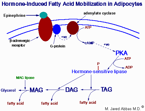
Model for the activation of hormone-sensitive lipase by epinephrine. Epinephrine binds its' receptor and leads to the activation of adenylate cyclase. The resultant increase in cAMP activates PKA which then phosphorylates and activates hormone-sensitive lipase. Hormone-sensitive lipase hydrolyzes fatty acids from triacylglycerols and diacylglycerols. The final fatty acid is released from monoacylglycerols through the action of monoacylglycerol lipase, an enzyme active in the absence of hormonal stimulation.

In contrast to the hormonal activation of adenylate cyclase and (subsequently) hormone-sensitive lipase in adipocytes, the mobilization of fat from adipose tissue is inhibited by numerous stimuli. The most significant inhibition is that exerted upon adenylate cyclase by insulin. When an individual is well fed state, insulin released from the pancreas prevents the inappropriate mobilization of stored fat. Instead, any excess fat and carbohydrate are incorporated into the triacylglycerol pool within adipose tissue.
back to the top
Reactions of Oxidation
Fatty acids must be activated in the cytoplasm before being oxidized in the mitochondria. Activation is catalyzed by fatty acyl-CoA ligase (also called acyl-CoA synthetase or thiokinase). The net result of this activation process is the consumption of 2 molar equivalents of ATP.

Fatty acid + ATP + CoA -------> Acyl-CoA + PPi + AMP

Oxidation of fatty acids occurs in the mitochondria. The transport of fatty acyl-CoA into the mitochondria is accomplished via an acyl-carnitine intermediate, which itself is generated by the action of carnitine acyltransferase I, an enzyme that resides in the outer mitochondrial membrane. The acyl-carnitine molecule then is transported into the mitochondria where carnitine acyltransferase II catalyzes the regeneration of the fatty acyl-CoA molecule.

Transport of fatty acids from the cytoplasm to the inner mitochondrial space for oxidation. Following activation to a fatty-CoA, the CoA is exchanged for carnitine by carnitine-palmitoyltransferase I. The fatty-carnitine is then transported to the inside of the mitochondrion where a reversal exchange takes place through the action of carnitine-palmitoyltransferase II. Once inside the mitochondrion the fatty-CoA is a substrate for the b-oxidation machinery.

The process of fatty acid oxidation is termed b-oxidation since it occurs through the sequential removal of 2-carbon units by oxidation at the b-carbon position of the fatty acyl-CoA molecule.
Each round of b-oxidation produces one mole of NADH, one mole of FADH2 and one mole of acetyl-CoA. The acetyl-CoA--- the end product of each round of b-oxidation--- then enters the TCA cycle, where it is further oxidized to CO2 with the concomitant generation of three moles of NADH, one mole of FADH2 and one mole of ATP. The NADH and FADH2 generated during the fat oxidation and acetyl-CoA oxidation in the TCA cycle then can enter the respiratory pathway for the production of ATP.
The oxidation of fatty acids yields significantly more energy per carbon atom than does the oxidation of carbohydrates. The net result of the oxidation of one mole of oleic acid (an 18-carbon fatty acid) will be 146 moles of ATP (2 mole equivalents are used during the activation of the fatty acid), as compared with 114 moles from an equivalent number of glucose carbon atoms.
back to the top
Alternative Oxidation Pathways
The majority of natural lipids contain an even number of carbon atoms. A small proportion that contain odd numbers; upon complete b-oxidation, these yield acetyl-CoA units plus a single mole of propionyl-CoA. The propionyl-CoA is converted, in an ATP-dependent pathway, to succinyl-CoA. The succinyl-CoA can then enter the TCA cycle for further oxidation.
The oxidation of unsaturated fatty acids is essentially the same process as for saturated fats, except when a double bond is encountered. In such a case, the bond is isomerized by a specific enoyl-CoA isomerase and oxidation continues. In the case of linoleate, the presence of the D-12 unsaturation results in the formation of a dienoyl-CoA during oxidation. This molecule is the substrate for an additional oxidizing enzyme, the NADPH requiring 2,4-dienoyl-CoA reductase.

Phytanic acid is a fatty acid present in the tissues of ruminants and in dairy products and is, therefore, an important dietary component of fatty acid intake. Because phytanic acid is methylated, it cannot act as a substrate for the first enzyme of the b-oxidation pathway (acyl-CoA dehydrogenase). An additional mitochondrial enzyme, a-hydroxylase, adds a hydroxyl group to the a-carbon of phytanic acid, which then serves as a substrate for the remainder of the normal oxidative enzymes. This process is termed a-oxidation.
back to the top
Regulation of Fatty Acid Metabolism
In order to understand how the synthesis and degradation of fats needs to be exquisitely regulated, one must consider the energy requirements of the organism as a whole. The blood is the carrier of triacylglycerols in the form of VLDLs and chylomicrons, fatty acids bound to albumin, amino acids, lactate, ketone bodies and glucose. The pancreas is the primary organ involved in sensing the organism's dietary and energetic states by monitoring glucose concentrations in the blood. Low blood glucose stimulates the secretion of glucagon, whereas, elevated blood glucose calls for the secretion of insulin.
The metabolism of fat is regulated by two distinct mechanisms. One is short-term regulation, which can come about through events such as substrate availability, allosteric effectors and/or enzyme modification. The other mechanism, long-term regulation, is achieved by alteration of the rate of enzyme synthesis and turn-over.
ACC is the rate-limiting (committed) step in fatty acid synthesis. This enzyme is activated by citrate and inhibited by palmitoyl-CoA and other long-chain fatty acyl-CoAs. ACC activity can also be affected by phosphorylation. For instance, glucagon-stimulated increases in PKA activity result in the phosphorylation of certain serine residues in ACC leading to decreased activity of the enzyme. By contrast, insulin leads to PKA-independent phosphorylation of ACC at sites distinct from glucagon, which bring about increased ACC activity. Both of these reaction chains are examples of short-term regulation.
Insulin, a product of the well-fed state, stimulates ACC and FAS synthesis, whereas starvation leads to a decrease in the synthesis of these enzymes. Adipose tissue levels of lipoprotein lipase also are increased by insulin and decreased by starvation. However, the effects of insulin and starvation on lipoprotein lipase in the heart are just the inverse of those in adipose tissue. This sensitivity allows the heart to absorb any available fatty acids in the blood in order to oxidize them for energy production. Starvation also leads to increases in the levels of cardiac enzymes of fatty acid oxidation, and to decreases in FAS and related enzymes of synthesis.
Adipose tissue contains hormone-sensitive lipase, which is activated by PKA-dependent phosphorylation; this activation increases the release of fatty acids into the blood. This in turn leads to the increased oxidation of fatty acids in other tissues such as muscle and liver. In the liver, the net result (due to increased acetyl-CoA levels) is the production of ketone bodies (see below). This would occur under conditions in which the carbohydrate stores and gluconeogenic precursors available in the liver are not sufficient to allow increased glucose production. The increased levels of fatty acid that become available in response to glucagon or epinephrine are assured of being completely oxidized, because PKA also phosphorylates ACC; the synthesis of fatty acid is thereby inhibited.
Insulin has the opposite effect to glucagon and epinephrine: it increases the synthesis of triacylglycerols (and glycogen). One of the many effects of insulin is to lower cAMP levels, which leads to increased dephosphorylation through the enhanced activity of protein phosphatases such as PP-1. With respect to fatty acid metabolism, this yields dephosphorylated and inactive hormone-sensitive lipase. Insulin also stimulates certain phosphorylation events. This occurs through activation of several cAMP-independent kinases, one of which phosphorylates and thereby stimulates the activity of ACC.
Fat metabolism can also be regulated by malonyl-CoA-mediated inhibition of carnitine acyltransferase I. Such regulation serves to prevent de novo synthesized fatty acids from entering the mitochondria and being oxidized.
back to the top
Clinical Significance of Fatty Acids
The majority of clinical problems related to fatty acid metabolism are associated with processes of oxidation.
These disorders fall into four main groups:
MCAD deficiency is the most common form of this disease. In the first years of life this deficiency will become apparent following a prolonged fasting period. Symptoms include vomiting, lethargy and frequently coma. Excessive urinary excretion of medium-chain dicarboxylic acids as well as their glycine and carnitine esters is diagnostic of this condition. In the case of this enzyme deficiency. taking care to avoid prolonged fasting is sufficient to prevent clinical problems.
  • 4. Refsum's Disease: Refsum's disease is a rare inherited disorder in which patients lack the mitochondrial a-oxidizing enzyme. As a consequence, they accumulate large quantities of phytanic acid in their tissues and serum. This leads to severe symptoms, including cerebellar ataxia, retinitis pigmentosa, nerve deafness and peripheral neuropathy. As expected, the restriction of dairy products and ruminant meat from the diet can ameliorate the symptoms of this disease.
back to the top
Ketogenesis
During high rates of fatty acid oxidation, primarily in the liver, large amounts of acetyl-CoA are generated. These exceed the capacity of the TCA cycle, and one result is the synthesis of ketone bodies, or ketogenesis. The ketone bodies are acetoacetate, b-hydroxybutyrate, and acetone.
The formation of acetoacetyl-CoA occurs by condensation of two moles of acetyl-CoA through a reversal of the thiolase catalyzed reaction of fat oxidation. Acetoacetyl-CoA and an additional acetyl-CoA are converted to b-hydroxy-b-methylglutaryl-CoA (HMG-CoA) by HMG-CoA synthase, an enzyme found in large amounts only in the liver. Some of the HMG-CoA leaves the mitochondria, where it is converted to mevalonate (the precursor for cholesterol synthesis) by HMG-CoA reductase. HMG-CoA in the mitochondria is converted to acetoacetate by the action of HMG-CoA lyase. Acetoacetate can undergo spontaneous decarboxylation to acetone, or be enzymatically converted to b-hydroxybutyrate through the action of b-hydroxybutyrate dehydrogenase. When the level of glycogen in the liver is high the production of b-hydroxybutyrate increases.
When carbohydrate utilization is low or deficient, the level of oxaloacetate will also be low, resulting in a reduced flux through the TCA cycle. This in turn leads to increased release of ketone bodies from the liver for use as fuel by other tissues. In early stages of starvation, when the last remnants of fat are oxidized, heart and skeletal muscle will consume primarily ketone bodies to preserve glucose for use by the brain. Acetoacetate and b-hydroxybutyrate, in particular, also serve as major substrates for the biosynthesis of neonatal cerebral lipids.
Ketone bodies are utilized by extrahepatic tissues through the conversion of b-hydroxybutyrate to acetoacetate and of acetoacetate to acetoacetyl-CoA. The first step involves the reversal of the b-hydroxybutyrate dehydrogenase reaction, and the second involves the action (shown below) of acetoacetate:succinyl-CoA transferase, also called ketoacyl-CoA-transferase.

Acetoacetate + Succinyl-CoA <------> Acetoacetyl-CoA + succinate

The latter enzyme is present in all tissues except the liver. Importantly, its absence allows the liver to produce ketone bodies but not to utilize them. This ensures that extrahepatic tissues have access to ketone bodies as a fuel source during prolonged starvation.
back to the top


Regulation of Ketogenesis
The fate of the products of fatty acid metabolism is determined by an individual's physiological status. Ketogenesis takes place primarily in the liver and may by affected by several factors:

  • 1. Control in the release of free fatty acids from adipose tissue directly affects the level of ketogenesis in the liver. This is, of course, substrate-level regulation.
  • 2. Once fats enter the liver, they have two distinct fates. They may be activated to acyl-CoAs and oxidized, or esterified to glycerol in the production of triacylglycerols. If the liver has sufficient supplies of glycerol-3-phosphate, most of the fats will be turned to the production of triacylglycerols.
  • 3. The generation of acetyl-CoA by oxidation of fats can be completely oxidized in the TCA cycle. Therefore, if the demand for ATP is high the fate of acetyl-CoA is likely to be further oxidation to CO2.
  • 4. The level of fat oxidation is regulated hormonally through phosphorylation of ACC, which may activate it (in response to glucagon) or inhibit it (in the case of insulin).

back to the top
Clinical Significance of Ketogenesis
The production of ketone bodies occurs at a relatively low rate during normal feeding and under conditions of normal physiological status. Normal physiological responses to carbohydrate shortages cause the liver to increase the production of ketone bodies from the acetyl-CoA generated from fatty acid oxidation. This allows the heart and skeletal muscles primarily to use ketone bodies for energy, thereby preserving the limited glucose for use by the brain.
The most significant disruption in the level of ketosis, leading to profound clinical manifestations, occurs in untreated insulin-dependent diabetes mellitus. This physiological state, diabetic ketoacidosis (DKA), results from a reduced supply of glucose (due to a significant decline in circulating insulin) and a concomitant increase in fatty acid oxidation (due to a concomitant increase in circulating glucagon). The increased production of acetyl-CoA leads to ketone body production that exceeds the ability of peripheral tissues to oxidize them. Ketone bodies are relatively strong acids (pKa around 3.5), and their increase lowers the pH of the blood. This acidification of the blood is dangerous chiefly because it impairs the ability of hemoglobin to bind oxygen.
back to the top
Back to Topics<<<<


This article has been modified by Dr. M. Javed Abbas.
If you have any comments please do not hesitate to sign my Guest Book.

20:42 21/12/2002