|
Back to Topics<<<< Definition: Blood filled sac or space formed by the widening or extension of the lumen of the artery, caused by the weakness of arterial wall that may be congenital, traumatic or pathological. Causes: Weakness in the arterial wall may be:
* Usually a penetrating wound to the artery * Occasionally a closed injury causing aneurysm of aorta * Rarely damage by fracture e.g. internal carotid artery * Arterio-venous aneurysm when acquired is due to the trauma * Arteriosclerosis – commonest, wall is weakened by atheroma or hypertension * Syphilis – Tunica media is weakened by endarteritis of vasa vasorum * Occasional complication subacute bacterial endocarditis – arterial wall weakness either because of an abscess formation or infected embolus rests there * In an artery traversing tubercular cavities in the lungs * In an artery located at the base of peptic ulcer 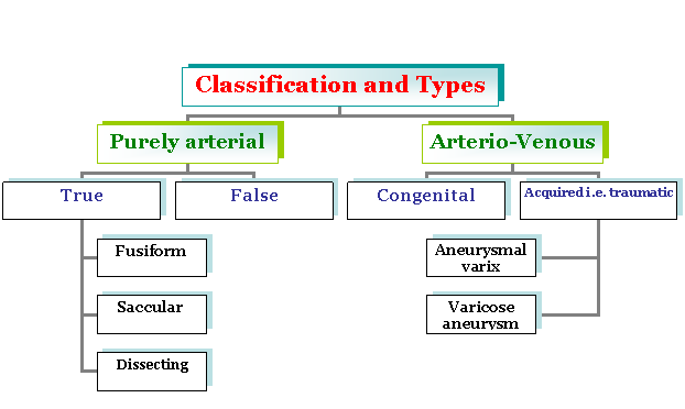 True Aneurysm: involves all the three layers of an artery. False aneurysm: intimal and medial layers are disrupted and dilation is lined by adventitia or sometimes by pervascular clot. When the dilation of an artery involves its whole circumference to give it spindle shaped enlargement, it is called fusiform aneurysm ( commonest type). When only small part of circumference of arterial wall stretches outwards as a rounded bulbous mass, it is called saccular aneurysm. These are usually traumatic. A penetrating wound in the artery doesn’t close spontaneously as retraction of intima keeps it patent. A pulsating hematoma develops, which is purified and enclosed by peripheral clot. Later, the saccular cavity becomes walled in by fibrous tissue and is partly lined by endothelium derived from intima. A dissecting aneurysm occurs when path of tunica intima ruptures (usually beneath atheromatous plaque) and blood is forced through the intima to collect and expand between inner and outer coats of tunica media (as if dissecting the arterial wall) In arteriovenous aneurysm (fistula), there is a communication between artery and a vein. This is due to:
Artery and vein communicate directly through a short wide channel – aneurismal varix Anastomoses can be indirect through an intermediate sac lying in the soft tissue – varicose aneurysm Etiology: For thoracic aneurysms the etiological factors are:
According to size Intrinsic:
Pain is localized upper thoracic region due to destruction of sympathetic plexus of aorta. Others are due to pressure effects on adjacent structures or due to distal arterial occlusion caused by the emboli that are formed in the aneurysmal sac and are carried distally. The effects of pressure on different organs and structures are as follows: Artery: distal pulse is smaller than that of the contralateral side and the pulse pressure in the distal vessel is reduced Gangrene: due to embolism Nerves: Altered sensation or paralysis Veins: Distal oedema Bones: May be eroded (e.g. vertebrae by aortic aneurysms) Tubes: Trachea, oesophagus may be compressed Skin: May be stretched and even necrosed Usually aortic thoracic aneurysm has juxtaposition to the 2nd, 3rd rib cartilage confluence to the sternum and clinically appears as swallowing pulsing elastic mass in this region. These are the prominent signs of aneurysm, sinuses and central vein distention, characteristic signs of thoracic aneurysm. Vena – Caval syndrome: Fistula occurs between aorta and inferior vena cava manifested in elevated pressure of various systems liner enlargement arteries. In non – operated patients, mortality rate is 37.5% after 3 years of diagnosis and 45% after 5 years of diagnosis. Abdominal aortic aneurysm should be defined as suprarenal and infrarenal one. Infrarenal type occurs in ľ cases. Copper was the first man who ligated the abdominal aneurysm in 1870. There is evidence of low velocity of blood and turbulence of blood flow in the lumen of artery. Main signs are sever acute pain, pulsing, elastic tumour mass in abdomen.
Abdominal syndrome: Belching, nausea and vomiting. Urologic syndrome: Dysuria and even hematuria. Ischio radical syndrome: Acute pain in back, radiating to leg because of compression of nerve plexus. Chronic ischemia: is caused by stagnation of blood by aneurysm. Complications:
Treatment: This is always surgical. Most commonly used current method is excision and artery graft i.e. reconstruction. The effected part of the aorta is resected and gap is replaced by prosthesis (e.g. Dacron graft) Dissecting aneurysm may be defined as separation of intima with elastic membrane. As a result of this, two luminal aorta appears after contrast investigation. This is life threatening form of aneurysm. Debaccy proposed complete classification of dissecting aneurysm:
Acute: acute form begins with the pain, shock and after 2hrs, patient dies Sub acute: its duration is 2-4 weeks Chronic: A chronic form continues for various months Main investigation method are ECG, CT scan, two dimensional echocardiography. Treatment is f choice. Penetrating injuries of the vessels are false after penetration. If there is connection between vessels, blood accumulates in the tissues (hematoma), then fibrous encapsulation occurs. In case of arteriovenous aneurysm, clinically elastic murmur occurs due to arterial venous shunt and there is also distinguishable sign. If we diminish the pulse, the pressure also diminishes within 10-15 beats. Treatment:
This article has been written by Dr. M. Javed Abbas. If you have any comments please do not hesitate to sign my Guest Book. 17:03 09/02/2003 |