|
Back to Topics<<<< Diverticula may be defined as outpouch from the wall of the hollow organ. There are two types of diverticulas: Real or congenital Diverticulas: Real or congenital diverticulas are usually uncommon and contain the elements from all the three layers of the hollow organ such as bowel e.g. Meckel’s Diverticulum. False or acquired diverticulas: False or acquired diverticulas are limited to mucous and submucous membrane and lack a proper muscular coat. As a matter of fact, false diverticulas can be referred as acquired herniation of the hollow organ wall when the mucous membrane due to some reasons protrudes through the muscular and serosal layers usually at a point where the blood vessels penetrate the hollow organ. The presentation of the diverticulas varies according to age. Incidence: at 40 years = 5% 60 Years = 30 % more than 60 years = 65% The main factor is high intraluminal pressure and elastic features of the bowel wall. About 90% diverticulas occur in the sigmoid colon. This must be described according to Lapalce law: where:
In sigmoid colon intraluminal radius least throughout the parameter of colon and intraluminal gradient pressure is higher. Anatomically the diverticulas are found in the weak areas of the colonic wall especially between mesenteric and antimesenteric teniae. Usual location of the diverticula is from antimesenteric teniae towards the mesenteric teniae. Here vasa recta penetrates muscular and serosal layer. These places must be considered as the weak areas. Diverticulitis appears as only 20 – 25% of the diverticular disease. Overall mortality rate is 5% after the first attack, which increases to as higher as 60% after the attacks of the diverticulitis. It is of two types:
In complicated type we have localized (epicolic) abscess formation or generalized peritonitis due to the perforation. It may be present with profuse haemorrhage (colonic) in 17% of the cases. Fistula formation occurs in 5% (Vesicolic, vaginocolic, enterocolic, colocutaneous). Clinical Signs:
Diverticulas result as a result of muscular incoordination and spasm, resulting in increased intraluminal pressure and segmentation. Excessive response to food, prostigmine and morphine is found in colonic motility studies, which is more apparent in symptomatic than in asymptomatic individuals. Histologically diverticulum consists of protrusion of mucous membrane covered with peritoneum. Thickening of circular muscular fibres teniae and intestine develops a saw-tooth appearance on barium enema. Diverticulas occur between muscle clefts making mucosal surface to appear trabaeculated. The elastic content of teniae coli is increased with controls. Bleeding occurs from the upper pole of diverticulas. In this region there is apparent thickening of intima of the vessels and thinning of media. Thickened intima tends to bleed after minute or little trauma. If the wall of colon is perforated, the contents may be localized or spread in the abdominal cavity. As a result we have abscess formation or generalized peritonitis. When there is abscess, tenderness of lower abdomen is distinguishable. Palpable fistulas are formed between the sigmoid portion of the colon and urinary bladder (uterus, skin etc.). Treatment: Uncomplicated diverticulitis:
If arteriography reveals the precise location of bleeding then intra-arterial injection of vasopressin may be used. This procedure facilitates the next surgical intervention, which includes segmental resection of colon. If there is no evidence of precise location of bleeding, then surgical intervention includes total colectomy. 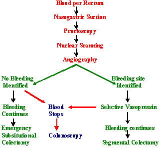 This article has been written by Dr. M. Javed Abbas. If you have any comments please do not hesitate to sign my Guest Book. 21:05 21/12/2002 |