Select From The Following for More Details
Back to Topics<<<<

Introduction to Cholesterol Metabolism

Cholesterol is an extremely important biological molecule that has roles in membrane structure as well as being a precursor for the synthesis of the steroid hormones and bile acids. Both dietary cholesterol and that synthesized de novo are transported through the circulation in lipoprotein particles. The same is true of cholesteryl esters, the form in which cholesterol is stored in cells.
The synthesis and utilization of cholesterol must be tightly regulated in order to prevent over-accumulation and abnormal deposition within the body. Of particular importance clinically is the abnormal deposition of cholesterol and cholesterol-rich lipoproteins in the coronary arteries. Such deposition, eventually leading to atherosclerosis, is the leading contributory factor in diseases of the coronary arteries.
Cholesterol
back to the top
Biosynthesis of Cholesterol
Slightly less than half of the cholesterol in the body derives from biosynthesis de novo. Biosynthesis in the liver accounts for approximately 10%, and in the intestines approximately 15%, of the amount produced each day. Cholesterol synthesis occurs in the cytoplasm and microsomes from the two-carbon acetate group of acetyl-CoA.

The process has five major steps:

  1. Acetyl-CoAs are converted to 3-hydroxy-3-methylglutaryl-CoA (HMG-CoA)
  2. HMG-CoA is converted to mevalonate
  3. Mevalonate is converted to the isoprene based molecule, isopentenyl pyrophosphate (IPP), with the concomitant loss of CO2
  4. IPP is converted to squalene
  5. Squalene is converted to cholesterol.
Pathway of cholesterol biosynthesis. Synthesis begins with the transport of acetyl-CoA ffrom the mitochondrion to the cytosol. The rate limiting step occurs at the 3-hydroxy-3-methylglutaryl-CoA (HMG-CoA) reducatse catalyzed step. The phosphorylation reactions are required to solubilize the isoprenoid intermediates in the pathway. Intermediates in the pathway are used for the synthesis of prenylated proteins, dolichol, coenzyme Q and the side chain of heme a.

The acetyl-CoA utilized for cholesterol biosynthesis is derived from an oxidation reaction (eg, fatty acids or pyruvate) in the mitochondria and is transported to the cytoplasm by the same process as that described for fatty acid synthesis. Acetyl-CoA can also be derived from cytoplasmic oxidation of ethanol by acetyl-CoA synthetase. All the reduction reactions of cholesterol biosynthesis use NADPH as a cofactor. The isoprenoid intermediates of cholesterol biosynthesis can be diverted to other synthesis reactions, such as those for dolichol (used in the synthesis of N-linked glycoproteins, coenzyme Q (of the oxidative phosphorylation) pathway or the side chain of heme a. Additionally, these intermediates are used in the lipid modification of some proteins.
Acetyl-CoA units are converted to mevalonate by a series of reactions that begins with the formation of HMG-CoA. Unlike the HMG-CoA formed during ketone body synthesis in the mitochondria, this form is synthesized in the cytoplasm. However, the pathway and the necessary enzymes are the same as those in the mitochondria. Two moles of acetyl-CoA are condensed in a reversal of the thiolase reaction, forming acetoacetyl-CoA. Acetoacetyl-CoA and a third mole of acetyl-CoA are converted to HMG-CoA by the action of HMG-CoA synthase. HMG-CoA is converted to mevalonate by HMG-CoA reductase (this enzyme is bound to the endoplasmic reticulum). HMG-CoA reductase absolutely requires NADPH as a cofactor and two moles of NADPH are consumed during the conversion of HMG-CoA to mevalonate.

The reaction catalyzed by HMG-CoA reductase is the rate limiting step of cholesterol biosynthesis, and this enzyme is subject to complex regulatory controls.

Mevalonate is then activated by three successive phosphorylations, yielding 5-pyrophosphomevalonate. In addition to activating mevalonate, the phosphorylations maintain its solubility, since otherwise it is insoluble in water. After phosphorylation, an ATP-dependent decarboxylation yields isopentenyl pyrophosphate, IPP, an activated isoprenoid molecule. Isopentenyl pyrophosphate is in equilibrium with its isomer, dimethylallyl pyrophosphate, DMPP. One molecule of IPP condenses with one molecule of DMPP to generate geranyl pyrophosphate, GPP. GPP further condenses with another IPP molecule to yield farnesyl pyrophosphate, FPP. Finally, the NADPH-requiring enzyme, squalene synthase catalyzes the head-to-tail condensation of two molecules of FPP, yielding squalene (squalene synthase also is tightly associated with the endoplasmic reticulum). Squalene undergoes a two step cyclization to yield lanosterol. The first reaction is catalyzed by squalene monooxygenase. This enzyme uses NADPH as a cofactor to introduce molecular oxygen as an epoxide at the 2,3 position of squalene. Through a series of 19 additional reactions, lanosterol is converted to cholesterol.
back to the top
Regulating Cholesterol Synthesis
Normal healthy adults synthesize cholesterol at a rate of approximately 1g/day and consume approximately 0.3g/day. A relatively constant level of cholesterol in the body (150 - 200 mg/dL) is maintained primarily by controlling the level of de novo synthesis. The level of cholesterol synthesis is regulated in part by the dietary intake of cholesterol. Cholesterol from both diet and synthesis is utilized in the formation of membranes and in the synthesis of the steroid hormones and bile acids (see below). The greatest proportion of cholesterol is used in bile acid synthesis.
The cellular supply of cholesterol is maintained at a steady level by three distinct mechanisms:
  1. Regulation of HMG-CoA reductase activity and levels
  2. Regulation of excess intracellular free cholesterol through the activity of acyl-CoA:cholesterol acyltransferase, ACAT
  3. Regulation of plasma cholesterol levels via LDL receptor-mediated uptake and HDL-mediated reverse transport.
Regulation of HMG-CoA reductase activity is the primary means for controlling the level of cholesterol biosynthesis. The enzyme is controlled by three distinct mechanisms: control of gene expression, rate of enzyme degradation and phosphorylation-dephosphorylation.
The first two control mechanisms are exerted by cholesterol itself. Cholesterol acts as a feed-back inhibitor of pre-existing HMG-CoA reductase as well as inducing rapid turn-over of the enzyme. In addition, when cholesterol is in excess the amount of mRNA for HMG-CoA reductase is reduced as a result of decreased expression of the gene. The exact mechanism for this cholesterol-induced regulation of gene activity is not known.
Regulation of HMG-CoA reductase through covalent modification occurs as a result of phosphorylation and dephosphorylation. The enzyme is most active in its unmodified form. Phosphorylation of the enzyme decreases its activity. HMG-CoA reductase is phosphorylated by AMP-regulated protein kinase, AMPRK (this is not the same as cAMP-dependent protein kinase, PKA). AMPRK itself is activated via phosphorylation. The phosphorylation of AMPRK is catalyzed by kinase kinase.

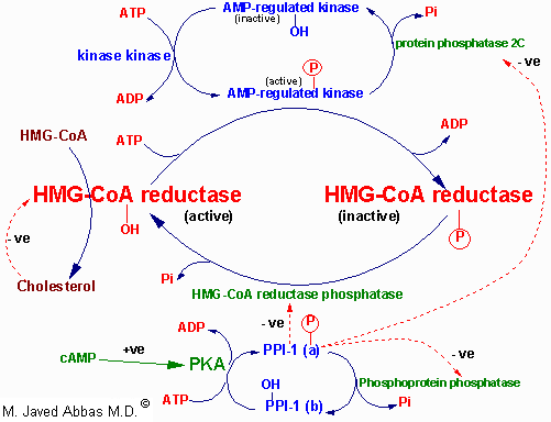
Regulation of HMG-CoA reductase by covalent modification. HMG-CoA reductase is most active in the dephosphorylated state. Phosphorylation is catalyzed by AMP-regulated protein kinase, AMPRK, (used to be termed reductase kinase), an enzyme whose activity is also regulated by phosphorylation. Phosphorylation of AMPRK is catalyzed by kinase kinase. Hormones such as glucagon and epinephrine negatively affect cholesterol biosynthesis by increasing the activity of the inhibitor of phosphoprotein phosphatase inhibitor-1, PPI-1. Conversely, insulin stimulates the removal of phosphates and, thereby, activates HMG-CoA reductase activity. Additional regulation of HMG-CoA reductase occurs through an inhibition of its' activity as well as of its' synthesis by elevation in intracellular cholesterol levels. The mechanism of this cholesterol induced inhibition is not fully understood.
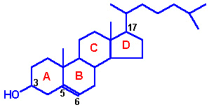
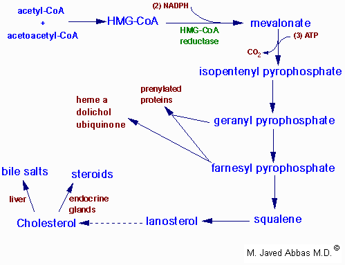
The activity of HMG-CoA reductase is further controlled by the cAMP signaling pathway. Increases in cAMP lead to activation of cAMP-dependent protein kinase, PKA. In the context of HMG-CoA reductase regulation, PKA phosphorylates phosphoprotein phosphatase inhibitor-1 (PPI-1) leading to an increase in its' activity. PPI-1 can inhibit the activity of numerous phosphatases including protein phosphatase 2C and HMG-CoA reductase phosphatase which remove phosphates from AMPRK and HMG-CoA reductase, respectively. This maintains AMPRK in the phosphorylated and active state, and HMG-CoA reductase in the phosphorylated and inactive state. As the stimulus leading to increased cAMP production is removed, the level of phosphorylations decreases and that of dephosphorylations increases. The net result is a return to a higher level of HMG-CoA reductase activity.
Since the intracellular level of cAMP is regulated by hormonal stimuli, regulation of cholesterol biosynthesis is hormonally controlled. Insulin leads to a decrease in cAMP, which in turn activates cholesterol synthesis. Alternatively, glucagon and epinephrine, which increase the level of cAMP, inhibit cholesterol synthesis.
The ability of insulin to stimulate, and glucagon to inhibit, HMG-CoA reductase activity is consistent with the effects of these hormones on other metabolic pathways. The basic function of these two hormones is to control the availability and delivery of energy to all cells of the body.
Long-term control of HMG-CoA reductase activity is exerted primarily through control over the synthesis and degradation of the enzyme. When levels of cholesterol are high, the level of expression of the HMG-CoA reductase gene is reduced. Conversely, reduced levels of cholesterol activate expression of the gene. Insulin also brings about long-term regulation of cholesterol metabolism by increasing the level of HMG-CoA reductase synthesis. The rate of HMG-CoA turn-over is also regulated by the supply of cholesterol. When cholesterol is abundant, the rate of HMG-CoA reductase degradation increases.
back to the top
The Utilization of Cholesterol
Cholesterol is transported in the plasma predominantly as cholesteryl esters associated with lipoproteins. Dietary cholesterol is transported from the small intestine to the liver within chylomicrons. Cholesterol synthesized by the liver, as well as any dietary cholesterol in the liver that exceeds hepatic needs, is transported in the serum within LDLs. The liver synthesizes VLDLs and these are converted to LDLs through the action of endothelial cell-associated lipoprotein lipase. Cholesterol found in plasma membranes can be extracted by HDLs and esterified by the HDL-associated enzyme LCAT. The cholesterol acquired from peripheral tissues by HDLs can then be transferred to VLDLs and LDLs via the action of cholesteryl ester transfer protein (apo-D) which is associated with HDLs. Reverse cholesterol transport allows peripheral cholesterol to be returned to the liver in LDLs. Ultimately, cholesterol is excreted in the bile as free cholesterol or as bile salts following conversion to bile acids in the liver.
back to the top
Bile Acids Synthesis and Utilization
The end products of cholesterol utilization are the bile acids, synthesized in the liver. Synthesis of bile acids is one of the predominant mechanisms for the excretion of excess cholesterol. However, the excretion of cholesterol in the form of bile acids is insufficient to compensate for an excess dietary intake of cholesterol.

Synthesis of the 2 primary bile acids, cholic acid and chenodeoxycholic acid. The reaction catalyzed by the 7a-hydroxylase is the rate limiting step in bile acid synthesis. Conversion of 7a-hydroxycholesterol to the bile acids requires several steps not shown in detail in this image. Only the relevant co-factors needed for the synthesis steps are shown.

The most abundant bile acids in human bile are chenodeoxycholic acid (45%) and cholic acid (31%). These are referred to as the primary bile acids. Within the intestines the primary bile acids are acted upon by bacteria and converted to the secondary bile acids, identified as deoxycholate (from cholate) and lithocholate (from chenodeoxycholate). Both primary and secondary bile acids are reabsorbed by the intestines and delivered back to the liver via the portal circulation.


Structure of the conjugated cholic acids

Within the liver the carboxyl group of primary and secondary bile acids is conjugated via an amide bond to either glycine or taurine before their being resecreted into the bile canaliculi. These conjugation reactions yield glycoconjugates and tauroconjugates, respectively. The bile canaliculi join with the bile ductules, which then form the bile ducts. Bile acids are carried from the liver through these ducts to the gallbladder, where they are stored for future use. The ultimate fate of bile acids is secretion into the intestine, where they aid in the emulsification of dietary lipids. In the gut the glycine and taurine residues are removed and the bile acids are either excreted (only a small percentage) or reabsorbed by the gut and returned to the liver. This process of secretion from the liver to the gallbladder, to the intestines and finally reabsorbtion is termed the enterohepatic circulation.
back to the top
Clinical Significance of Bile Acid Synthesis

Bile acids perform four physiologically significant functions:
  1. their synthesis and subsequent excretion in the feces represent the only significant mechanism for the elimination of excess cholesterol.
  2. bile acids and phospholipids solubilize cholesterol in the bile, thereby preventing the precipitation of cholesterol in the gallbladder.
  3. they facilitate the digestion of dietary triacylglycerols by acting as emulsifying agents that render fats accessible to pancreatic lipases.
  4. they facilitate the intestinal absorption of fat-soluble vitamins.
back to the top
Back to Topics<<<<


This article has been modified by Dr. M. Javed Abbas.
If you have any comments please do not hesitate to sign my Guest Book.

20:43 21/12/2002