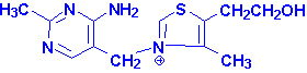
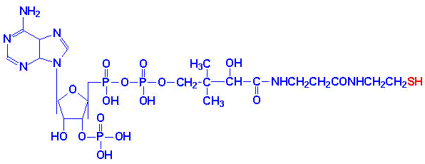
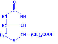
Select From The Following for More Details
Back to Topics<<<<
Water Soluble Vitamins Fat Soluble Vitamins


Thiamin

Thiamin structure

Thiamin is also known as vitamin B1 . Thiamin is derived from a substituted pyrimidine and a thiazole which are coupled by a methylene bridge. Thiamin is rapidly converted to its active form, thiamin pyrophosphate, TPP, in the brain and liver by a specific enzymes, thiamin diphosphotransferase.
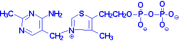
Thiamin pyrophosphate

TPP is necessary as a cofactor for the pyruvate and a-ketoglutarate dehydrogenase catalyzed reactions as well as the transketolase catalyzed reactions of the pentose phosphate pathway. A deficiency in thiamin intake leads to a severely reduced capacity of cells to generate energy as a result of its role in these reactions.
The dietary requirement for thiamin is proportional to the caloric intake of the diet and ranges from 1.0 - 1.5 mg/day for normal adults. If the carbohydrate content of the diet is excessive then an in thiamin intake will be required.
back to the top

Clinical Significances of Thiamin Deficiency

The earliest symptoms of thiamin deficiency include constipation, appetite suppression, nausea as well as mental depression, peripheral neuropathy and fatigue. Chronic thiamin deficiency leads to more severe neurological symptoms including ataxia, mental confusion and loss of eye coordination. Other clinical symptoms of prolonged thiamin deficiency are related to cardiovascular and musculature defects.
The severe thiamin deficiency disease known as Beriberi, is the result of a diet that is carbohydrate rich and thiamin deficient. An additional thiamin deficiency related disease is known as Wernicke-Korsakoff syndrome. This disease is most commonly found in chronic alcoholics due to their poor dietetic lifestyles.
back to the top

Riboflavin

Riboflavin structure

Riboflavin is also known as vitamin B2. Riboflavin is the precursor for the coenzymes, flavin mononucleotide (FMN) and flavin adenine dinucleotide (FAD). The enzymes that require FMN or FAD as cofactors are termed flavoproteins. Several flavoproteins also contain metal ions and are termed metalloflavoproteins. Both classes of enzymes are involved in a wide range of redox reactions, e.g. succinate dehydrogenase and xanthine oxidase. During the course of the enzymatic reactions involving the flavoproteins the reduced forms of FMN and FAD are formed, FMNH2 and FADH2, respectively.
Structure of FAD
nitrogens 1 & 5 carry hydrogens in FADH2
The normal daily requirement for riboflavin is 1.2 - 1.7 mg/day for normal adults.
back to the top

Clinical Significances of Flavin Deficiency

Riboflavin deficiencies are rare in the United States due to the presence of adequate amounts of the vitamin in eggs, milk, meat and cereals. Riboflavin deficiency is often seen in chronic alcoholics due to their poor dietetic habits.
Symptoms associated with riboflavin deficiency include, glossitis, seborrhea, angular stomatitis, cheilosis and photophobia. Riboflavin decomposes when exposed to visible light. This characteristic can lead to riboflavin deficiencies in newborns treated for hyperbilirubinemia by phototherapy.
back to the top
Niacin

Nicotinamide Nicotinic Acid
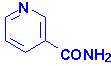
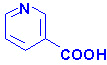
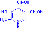
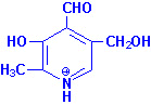
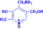
Niacin (nicotinic acid and nicotinamide) is also known as vitamin B3. Both nicotinic acid and nicotinamide can serve as the dietary source of vitamin B3. Niacin is required for 
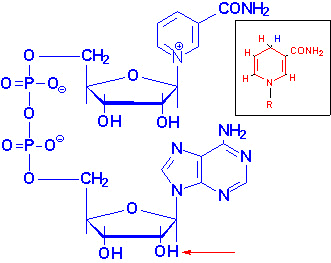
the synthesis of the active forms of vitamin B3, nicotinamide adenine dinucleotide (NAD+) and nicotinamide adenine dinucleotide phosphate (NADP+). Both NAD+ and NADP+ function as cofactors for numerous dehydrogenase, e.g., lactate and malate dehydrogenases.

Structure of NAD+
NADH is shown in the box insert.
The -OH phosphorylated in NADP+ is indicated by the red arrow.

Niacin is not a true vitamin in the strictest definition since it can be derived from the amino acid tryptophan. However, the ability to utilize tryptophan for niacin synthesis is inefficient (60 mg of tryptophan are required to synthesize 1 mg of niacin). Also, synthesis of niacin from tryptophan requires vitamins B1, B2 and B6 which would be limiting in themselves on a marginal diet.
The recommended daily requirement for niacin is 13 - 19 niacin equivalents (NE) per day for a normal adult. One NE is equivalent to 1 mg of free niacin).
back to the top
Clinical Significances of Niacin and Nicotinic Acid
A diet deficient in niacin (as well as tryptophan) leads to glossitis of the tongue, dermatitis, weight loss, diarrhea, depression and dementia. The severe symptoms, depression, dermatitis and diarrhea, are associated with the condition known as pellagra. Several physiological conditions (e.g. Hartnup disease and malignant carcinoid syndrome) as well as certain drug therapies (e.g. isoniazid) can lead to niacin deficiency. In Hartnup disease tryptophan absorption is impaired and in malignant carcinoid syndrome tryptophan metabolism is altered resulting in excess serotonin synthesis. Isoniazid (the hydrazide derivative of isonicotinic acid) is the primary drug for chemotherapy of tuberculosis.
Nicotinic acid (but not nicotinamide) when administered in pharmacological doses of 2 - 4 g/day lowers plasma cholesterol levels and has been shown to be a useful therapeutic for hypercholesterolemia. The major action of nicotinic acid in this capacity is a reduction in fatty acid mobilization from adipose tissue. Although nicotinic acid therapy lowers blood cholesterol it also causes a depletion of glycogen stores and fat reserves in skeletal and cardiac muscle. Additionally, there is an elevation in blood glucose and uric acid production. For these reasons nicotinic acid therapy is not recommended for diabetics or persons who suffer from gout.
back to the top
Pantothenic Acid
Pantothenic Acid
Pantothenic acid is also known as vitamin B5. Pantothenic acid is formed from b-alanine and pantoic acid. Pantothenate is required for synthesis of coenzyme A, CoA and is a component of the acyl carrier protein (ACP) domain of fatty acid synthase. Pantothenate is, therefore, required for the metabolism of carbohydrate via the TCA cycle and all fats and proteins. At least 70 enzymes have been identified as requiring CoA or ACP derivatives for their function.
Deficiency of pantothenic acid is extremely rare due to its widespread distribution in whole grain cereals, legumes and meat. Symptoms of pantothenate deficiency are difficult to assess since they are subtle and resemble those of other B vitamin deficiencies.

Coenzyme A

back to the top
Vitamin B6
Pyridoxine Pyridoxal Pyridoxamine

Pyridoxal, pyridoxamine and pyridoxine are collectively known as vitamin B6. All three compounds are efficiently converted to the biologically active form of vitamin B6, pyridoxal phosphate. This conversion is catalyzed by the ATP requiring enzyme, pyridoxal kinase.

Pyridoxal Phosphate
Pyridoxal phosphate functions as a cofactor in enzymes involved in transamination reactions required for the synthesis and catabolism of the amino acids as well as in glycogenolysis as a cofactor for glycogen phosphorylase. The requirement for vitamin B6 in the diet is proportional to the level of protein consumption ranging from 1.4 - 2.0 mg/day for a normal adult. During pregnancy and lactation the requirement for vitamin B6 increases approximately 0.6 mg/day.
Deficiencies of vitamin B6 are rare and usually are related to an overall deficiency of all the B-complex vitamins. Isoniazid (see niacin deficiencies above) and penicillamine (used to treat rheumatoid arthritis and cystinurias) are two drugs that complex with pyridoxal and pyridoxal phosphate resulting in a deficiency in this vitamin.
back to the top
Biotin
Biotin
Biotin is the cofactor required of enzymes that are involved in carboxylation reactions, e.g. acetyl-CoA carboxylase and pyruvate carboxylase. Biotin is found in numerous foods and also is synthesized by intestinal bacteria and as such deficiencies of the vitamin are rare. Deficiencies are generally seen only after long antibiotic therapies which deplete the intestinal fauna or following excessive consumption of raw eggs. The latter is due to the affinity of the egg white protein, avidin, for biotin preventing intestinal absorption of the biotin.
back to the top
Cobalamin
Cobalamin is more commonly known as vitamin B12. Vitamin B12 is composed of a complex tetrapyrrol ring structure (corrin ring) and a cobalt ion in the center. Vitamin B12 is synthesized exclusively by microorganisms and is found in the liver of animals bound to protein as methycobalamin or 5'-deoxyadenosylcobalamin. The vitamin must be hydrolyzed from protein in order to be active. Hydrolysis occurs in the stomach by gastric acids or the intestines by trypsin digestion following consumption of animal meat. The vitamin is then bound by intrinsic factor, a protein secreted by parietal cells of the stomach, and carried to the ileum where it is absorbed. Following absorption the vitamin is transported to the liver in the blood bound to transcobalamin II.
There are only two clinically significant reactions in the body that require vitamin B12 as a cofactor. During the catabolism of fatty acids with an odd number of carbon atoms and the amino acids valine, isoleucine and threonine the resultant propionyl-CoA is converted to succinyl-CoA for oxidation in the TCA cycle. One of the enzymes in this pathway, methylmalonyl-CoA mutase, requires vitamin B12 as a cofactor in the conversion of methylmalonyl-CoA to succinyl-CoA. The 5'-deoxyadenosine derivative of cobalamin is required for this reaction.
The second reaction requiring vitamin B12 catalyzes the conversion of homocysteine to methionine and is catalyzed by methionine synthase. This reaction results in the transfer of the methyl group from N5-methyltetrahydrofolate to hydroxycobalamin generating tetrahydrofolate (THF) and methylcobalamin during the process of the conversion.
Deoxyadenosylcobalamin
back to the top
Clinical Significances of B12 Deficiency
The liver can store up to six years worth of vitamin B12, hence deficiencies in this vitamin are rare. Pernicious anemia is a megaloblastic anemia resulting from vitamin B12 deficiency that develops as a result a lack of intrinsic factor in the stomach leading to malabsorption of the vitamin. The anemia results from impaired DNA synthesis due to a block in purine and thymidine biosynthesis. The block in nucleotide biosynthesis is a 
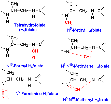
consequence of the effect of vitamin B12 on folate metabolism. When vitamin B12 is deficient essentially all of the folate becomes trapped as the N5-methylTHF derivative as a result of the loss of functional methionine synthase. This trapping prevents the synthesis of other THF derivatives required for the purine and thymidine nucleotide biosynthesis pathways.
Neurological complications also are associated with vitamin B12 deficiency and result from a progressive demyelination of nerve cells. The demyelination is thought to result from the increase in methylmalonyl-CoA that result from vitamin B12 deficiency. Methylmalonyl-CoA is a competitive inhibitor of malonyl-CoA in fatty acid biosynthesis as well as being able to substitute for malonyl-CoA in any fatty acid biosynthesis that may occur. Since the myelin sheath is in continual flux the methylmalonyl-CoA-induced inhibition of fatty acid synthesis results in the eventual destruction of the sheath. The incorporation methylmalonyl-CoA into fatty acid biosynthesis results in branched-chain fatty acids being produced that may severely alter the architecture of the normal membrane structure of nerve cells
back to the top
Folic Acid

Folic Acid
positions 7 & 8 carry hydrogens in dihydrofolate (DHF)
positions 5-8 carry hydrogens in tetrahydrofolate (THF)

Folic acid is a conjugated molecule consisting of a pteridine ring structure linked to para-aminobenzoic acid (PABA) that forms pteroic acid. Folic acid itself is then generated through the conjugation of glutamic acid residues to pteroic acid. Folic acid is obtained primarily from yeasts and leafy vegetables as well as animal liver. Animal cannot synthesize PABA nor attach glutamate residues to pteroic acid, thus, requiring folate intake in the diet.
When stored in the liver or ingested folic acid exists in a polyglutamate form. Intestinal mucosal cells remove some of the glutamate residues through the action of the lysosomal enzyme, conjugase. The removal of glutamate residues makes folate less negatively charged (from the polyglutamic acids) and therefore more capable of passing through the basal lamenal membrane of the epithelial cells of the intestine and into the bloodstream. Folic acid is reduced within cells (principally the liver where it is stored) to tetrahydrofolate (THF also H4folate) through the action of dihydrofolate reductase (DHFR), an NADPH-requiring enzyme.
The function of THF derivatives is to carry and transfer various forms of one carbon units during biosynthetic reactions. The one carbon units are either methyl, methylene, methenyl, formyl or formimino groups.

Active center of tetrahydrofolate (THF). Note that the N5 position is the site of attachment of methyl groups, the N10 the site for attachment of formyl and formimino groups and that both N5 and N10 bridge the methylene and methenyl groups.
These one carbon transfer reactions are required in the biosynthesis of serine, methionine, glycine, choline and the purine nucleotides and dTMP.
The ability to acquire choline and amino acids from the diet and to salvage the purine nucleotides makes the role of N5,N10-methylene-THF in dTMP synthesis the most metabolically significant function for this vitamin. The role of vitamin B12 and N5-methyl-THF in the conversion of homocysteine to methionine also can have a significant impact on the ability of cells to regenerate needed THF.
back to the top
Clinical Significance of Folate Deficiency
Folate deficiency results in complications nearly identical to those described for vitamin B12 deficiency. The most pronounced effect of folate deficiency on cellular processes is upon DNA synthesis. This is due to an impairment in dTMP synthesis which leads to cell cycle arrest in S-phase of rapidly proliferating cells, in particular hematopoietic cells. The result is megaloblastic anemia as for vitamin B12 deficiency. The inability to synthesize DNA during erythrocyte maturation leads to abnormally large erythrocytes termed macrocytic anemia.
Folate deficiencies are rare due to the adequate presence of folate in food. Poor dietary habits as those of chronic alcoholics can lead to folate deficiency. The predominant causes of folate deficiency in non-alcoholics are impaired absorption or metabolism or an increased demand for the vitamin. The predominant condition requiring an increase in the daily intake of folate is pregnancy. This is due to an increased number of rapidly proliferating cells present in the blood. The need for folate will nearly double by the third trimester of pregnancy. Certain drugs such as anticonvulsants and oral contraceptives can impair the absorption of folate. Anticonvulsants also increase the rate of folate metabolism.
back to the top
Ascorbic Acid
Ascorbic Acid
Ascorbic acid is more commonly known as vitamin C. Ascorbic acid is derived from glucose via the uronic acid pathway. The enzyme L-gulonolactone oxidase responsible for the conversion of gulonolactone to ascorbic acid is absent in primates making ascorbic acid required in the diet.
The active form of vitamin C is ascorbate acid itself. The main function of ascorbate is as a reducing agent in a number of different reactions. Vitamin C has the potential to reduce cytochromes a and c of the respiratory chain as well as molecular oxygen. The most important reaction requiring ascorbate as a cofactor is the hydroxylation of proline residues in collagen. Vitamin C is, therefore, required for the maintenance of normal connective tissue as well as for wound healing since synthesis of connective tissue is the first event in wound tissue remodeling. Vitamin C also is necessary for bone remodeling due to the presence of collagen in the organic matrix of bones.
Several other metabolic reactions require vitamin C as a cofactor. These include the catabolism of tyrosine and the synthesis of epinephrine from tyrosine and the synthesis of the bile acids. It is also believed that vitamin C is involved in the process of steroidogenesis since the adrenal cortex contains high levels of vitamin C which are depleted upon adrenocorticotropic hormone (ACTH) stimulation of the gland.
Deficiency in vitamin C leads to the disease scurvy due to the role of the vitamin in the post-translational modification of collagens. Scurvy is characterized by easily bruised skin, muscle fatigue, soft swollen gums, decreased wound healing and hemorrhaging, osteoporosis, and anemia. Vitamin C is readily absorbed and so the primary cause of vitamin C deficiency is poor diet and/or an increased requirement. The primary physiological state leading to an increased requirement for vitamin C is severe stress (or trauma). This is due to a rapid depletion in the adrenal stores of the vitamin. The reason for the decrease in adrenal vitamin C levels is unclear but may be due either to redistribution of the vitamin to areas that need it or an overall increased utilization.
back to the top
Vitamin A
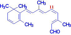
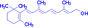
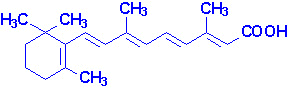
Vitamin A consists of three biologically active molecules, retinol, retinal (retinaldehyde) and retinoic acid.

All-trans-retinal 11-cis-retinal

Retinol Retinoic Acid

Each of these compounds are derived from the plant precursor molecule, b-carotene (a member of a family of molecules known as carotenoids). Beta-carotene, which consists of two molecules of retinal linked at their aldehyde ends, is also referred to as the provitamin form of vitamin A.
Ingested b-carotene is cleaved in the lumen of the intestine by b-carotene dioxygenase to yield retinal. Retinal is reduced to retinol by retinaldehyde reductase, an NADPH requiring enzyme within the intestines. Retinol is esterified to palmitic acid and delivered to the blood via chylomicrons. The uptake of chylomicron remnants by the liver results in delivery of retinol to this organ for storage as a lipid ester within lipocytes. Transport of retinol from the liver to extrahepatic tissues occurs by binding of hydrolyzed retinol to aporetinol binding protein (RBP). the retinol-RBP complex is then transported to the cell surface within the Golgi and secreted. Within extrahepatic tissues retinol is bound to cellular retinol binding protein (CRBP). Plasma transport of retinoic acid is accomplished by binding to albumin.
back to the top

Gene Control Exerted by Retinol and Retinoic Acid

Within cells both retinol and retinoic acid bind to specific receptor proteins. Following binding, the receptor-vitamin complex interacts with specific sequences in several genes involved in growth and differentiation and affects expression of these genes. In this capacity retinol and retinoic acid are considered hormones of the steroid/thyroid hormone superfamily of proteins. Vitamin D also acts in a similar capacity. Several genes whose patterns of expression are altered by retinoic acid are involved in the earliest processes of embryogenesis including the differentiation of the three germ layers, organogenesis and limb development.
back to the top
Vision and the Role of Vitamin A
Photoreception in the eye is the function of two specialized cell types located in the retina; the rod and cone cells. Both rod and cone cells contain a photoreceptor pigment in their membranes. The photosensitive compound of most mammalian eyes is a protein called opsin to which is covalently coupled an aldehyde of vitamin A. The opsin of rod cells is called scotopsin. The photoreceptor of rod cells is specifically called rhodopsin or visual purple. This compound is a complex between scotopsin and the 11-cis-retinal (also called 11-cis-retinene) form of vitamin A. Rhodopsin is a serpentine receptor imbedded in the membrane of the rod cell. Coupling of 11-cis-retinal occurs at three of the transmembrane domains of rhodopsin. Intracellularly, rhodopsin is coupled to a specific G-protein called transducin.
When the rhodopsin is exposed to light it is bleached releasing the 11-cis-retinal from opsin. Absorption of photons by 11-cis-retinal triggers a series of conformational changes on the way to conversion all-trans-retinal. One important conformational intermediate is metarhodopsin II. The release of opsin results in a conformational change in the photoreceptor. This conformational change activates transducin, leading to an increased GTP-binding by the a-subunit of transducin. Binding of GTP releases the a-subunit from the inhibitory b- and g-subunits. The GTP-activated a-subunit in turn activates an associated phosphodiesterase; an enzyme that hydrolyzes cyclic-GMP (cGMP) to GMP. Cyclic GMP is required to maintain the Na+ channels of the rod cell in the open conformation. The drop in cGMP concentration results in complete closure of the Na+ channels. Metarhodopsin II appears to be responsible for initiating the closure of the channels. The closing of the channels leads to hyperpolarization of the rod cell with concomitant propagation of nerve impulses to the brain.
back to the top
Additional Role of Retinol
Retinol also functions in the synthesis of certain glycoproteins and mucopolysaccharides necessary for mucous production and normal growth regulation. This is accomplished by phosphorylation of retinol to retinyl phosphate which then functions similarly to dolichol phosphate.
back top the top
Clinical Significances of Vitamin A Deficiency
Vitamin A is stored in the liver and deficiency of the vitamin occurs only after prolonged lack of dietary intake. The earliest symptoms of vitamin A deficiency are night blindness. Additional early symptoms include follicular hyperkeratinosis, increased susceptibility to infection and cancer and anemia equivalent to iron deficient anemia. Prolonged lack of vitamin A leads to deterioration of the eye tissue through progressive keratinization of the cornea, a condition known as xerophthalmia.
The increased risk of cancer in vitamin deficiency is thought to be the result of a depletion in b-carotene. Beta-carotene is a very effective antioxidant and is suspected to reduce the risk of cancers known to be initiated by the production of free radicals. Of particular interest is the potential benefit of increased b-carotene intake to reduce the risk of lung cancer in smokers. However, caution needs to be taken when increasing the intake of any of the lipid soluble vitamins. Excess accumulation of vitamin A in the liver can lead to toxicity which manifests as bone pain, hepatosplenomegaly, nausea and diarrhea.
back to the top
Vitamin D
Vitamin D is a steroid hormone that functions to regulate specific gene expression following interaction with its intracellular receptor. The biologically active form of the hormone is 1,25-dihydroxy vitamin D3 (1,25-(OH)2D3, also termed calcitriol). Calcitriol functions primarily to regulate calcium and phosphorous homeostasis.

Ergosterol Vitamin D2

7-Dehydrocholesterol Vitamin D3

Active calcitriol is derived from ergosterol (produced in plants) and from 7-dehydrocholesterol (produced in the skin). Ergocalciferol (vitamin D2) is formed by uv irradiation of ergosterol. In the skin 7-dehydrocholesterol is converted to cholecalciferol (vitamin D3) following uv irradiation.
Vitamin D2 and D3 are processed to D2-calcitriol and D3-calcitriol, respectively, by the same enzymatic pathways in the body. Cholecalciferol (or egrocalciferol) are absorbed from the intestine and transported to the liver bound to a specific vitamin D-binding protein. In the liver cholecalciferol is hydroxylated at the 25 position by a specific D3-25-hydroxylase generating 25-hydroxy-D3 [25-(OH)D3] which is the major circulating form of vitamin D. Conversion of 25-(OH)D3 to its biologically active form, calcitriol, occurs through the activity of a specific D3-1-hydroxylase present in the proximal convoluted tubules of the kidneys, and in bone and placenta. 25-(OH)D3 can also be hydroxylated at the 24 position by a specific D3-24-hydroxylase in the kidneys, intestine, placenta and cartilage.

25-hydroxyvitamin D3 1,25-dihydroxyvitamin D3

Calcitriol functions in concert with parathyroid hormone (PTH) and calcitonin to regulate serum calcium and phosphorous levels. PTH is released in response to low serum calcium and induces the production of calcitriol. In contrast, reduced levels of PTH stimulate synthesis of the inactive 24,25-(OH)2D3. In the intestinal epithelium, calcitriol functions as a steroid hormone in inducing the expression of calbindinD28K, a protein involved in intestinal calcium absorption. The increased absorption of calcium ions requires concomitant absorption of a negatively charged counter ion to maintain electrical neutrality. The predominant counter ion is Pi. When plasma calcium levels fall the major sites of action of calcitriol and PTH are bone where they stimulate bone resorption and the kidneys where they inhibit calcium excretion by stimulating reabsorption by the distal tubules. The role of calcitonin in calcium homeostasis is to decrease elevated serum calcium levels by inhibiting bone resorption.
back to the top
Clinical Significance of Vitamin D Deficiency
As a result of the addition of vitamin D to milk, deficiencies in this vitamin are rare in this country. The main symptom of vitamin D deficiency in children is rickets and in adults is osteomalacia. Rickets is characterized improper mineralization during the development of the bones resulting in soft bones. Osteomalacia is characterized by demineralization of previously formed bone leading to increased softness and susceptibility to fracture.
back to the top
Vitamin E
a-Tocopherol
Vitamin E is a mixture of several related compounds known as tocopherols. The a-tocopherol molecule is the most potent of the tocopherols. Vitamin E is absorbed from the intestines packaged in chylomicrons. It is delivered to the tissues via chylomicron transport and then to the liver through chylomicron remnant uptake. The liver can export vitamin E in VLDLs. Due to its lipophilic nature, vitamin E accumulates in cellular membranes, fat deposits and other circulating lipoproteins. The major site of vitamin E storage is in adipose tissue.
The major function of vitamin E is to act as a natural antioxidant by scavenging free radicals and molecular oxygen. In particular vitamin E is important for preventing peroxidation of polyunsaturated membrane fatty acids. The vitamins E and C are interrelated in their antioxidant capabilities. Active a-tocopherol can be regenerated by interaction with vitamin C following scavenge of a peroxy free radical. Alternatively, a-tocopherol can scavenge two peroxy free radicals and then be conjugated to glucuronate for excretion in the bile.
back to the top
Clinical significances of Vitamin E Deficiency
No major disease states have been found to be associated with vitamin E deficiency due to adequate levels in the average American diet. The major symptom of vitamin E deficiency in humans is an increase in red blood cell fragility. Since vitamin E is absorbed from the intestines in chylomicrons, any fat malabsorption diseases can lead to deficiencies in vitamin E intake. Neurological disorders have been associated with vitamin E deficiencies associated with fat malabsorptive disorders. Increased intake of vitamin E is recommended in premature infants fed formulas that are low in the vitamin as well as in persons consuming a diet high in polyunsaturated fatty acids. Polyunsaturated fatty acids tend to form free radicals upon exposure to oxygen and this may lead to an increased risk of certain cancers.
back to the top
Vitamin K
The K vitamins exist naturally as K1 (phylloquinone) in green vegetables and K2 (menaquinone) produced by intestinal bacteria and K3 is synthetic menadione. When administered, vitamin K3 is alkylated to one of the vitamin K2 forms of menaquinone.

Vitamin K1
Vitamin K2
"n" can be 6, 7 or 9 isoprenoid groups
Vitamin K3
The major function of the K vitamins is in the maintenance of normal levels of the blood clotting proteins, factors II, VII, IX, X and protein C and protein S, which are synthesized in the liver as inactive precursor proteins. Conversion from inactive to active clotting factor requires a posttranslational modification of specific glutamate (E) residues. This modification is a carboxylation and the enzyme responsible requires vitamin K as a cofactor. The resultant modified E residues are g-carboxyglutamate (gla). This process is most clearly understood for factor II, also called preprothrombin. Prothrombin is modified preprothrombin. The gla residues are effective calcium ion chelators. Upon chelation of calcium, prothrombin interacts with phospholipids in membranes and is proteolysed to thrombin through the action of activated factor X (Xa).
During the carboxylation reaction reduced hydroquinone form of vitamin K is converted to a 2,3-epoxide form. The regeneration of the hydroquinone form requires an uncharacterized reductase. This latter reaction is the site of action of the dicumarol based anticoagulants such as warfarin.
back to the top
Clinical significance of Vitamin K Deficiency
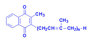
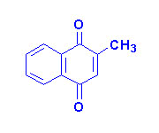
Naturally occurring vitamin K is absorbed from the intestines only in the presence of bile salts and other lipids through interaction with chylomicrons. Therefore, fat malabsorptive diseases can result in vitamin K deficiency. The synthetic vitamin K3 is water soluble and absorbed irrespective of the presence of intestinal lipids and bile. Since the vitamin K2 form is synthesized by intestinal bacteria, deficiency of the vitamin in adults is rare. However, long term antibiotic treatment can lead to deficiency in adults. The intestine of newborn infants is sterile, therefore, vitamin K deficiency in infants is possible if lacking from the early diet. The primary symptom of a deficiency in infants is a hemorrhagic syndrome.
back to the top
Back to Topics<<<<


This article has been modified by Dr. M. Javed Abbas.
If you have any comments please do not hesitate to sign my Guest Book.

20:38 21/12/2002