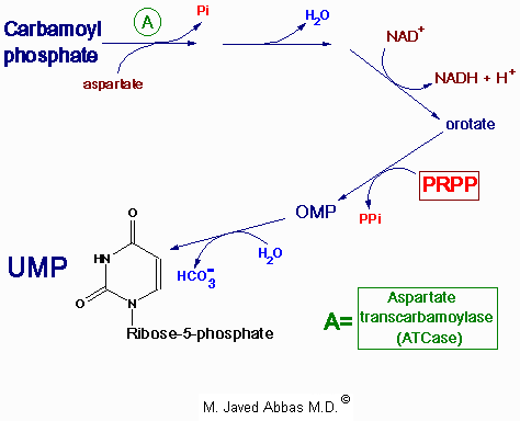
Select From The Following for More Details
Back to Topics<<<<

Introduction
The metabolic requirements for the nucleotides and their cognate bases can be met by both dietary intake or synthesis de novo from low molecular weight precursors. Indeed, the ability to salvage nucleotides from sources within the body alleviates any nutritional requirement for nucleotides, thus the purine and pyrimidine bases are not required in the diet. The salvage pathways are a major source of nucleotides for synthesis of DNA, RNA and enzyme co-factors.
Extracellular hydrolysis of ingested nucleic acids occurs through the concerted actions of endonucleases, phosphodiesterases and nucleoside phosphorylases. Endonucleases degrade DNA and RNA at internal sites leading to the production of oligonucleotides. Oligonucleotides are further digested by phosphodiesterases that act from the ends inward yielding free nucleosides. The bases are hydrolyzed from nucleosides by the action of phosphorylases that yield ribose-1-phosphate and free bases. If the nucleosides and/or bases are not re-utilized the purine bases are further degraded to uric acid and the pyrimidines to b-aminoiosobutyrate, NH3 and CO2.
Both the salvage and de novo synthesis pathways of purine and pyrimidine biosynthesis lead to production of nucleoside-5'-phosphates through the utilization of an activated sugar intermediate and a class of enzymes called phosphoribosyltransferases. The activated sugar used is 5-phosphoribosyl-1-pyrophosphate, PRPP. PRPP is generated by the action of PRPP synthetase and requires energy in the form of ATP as shown:

ribose-5-phosphate + ATP -------> PRPP + AMP

Note that this reaction releases AMP. Therefore, 2 high energy phosphate equivalents are consumed during the reaction.
back to the top
Purine Nucleotide Biosynthesis
The major site of purine synthesis is in the liver. Synthesis of the purine nucleotides begins with PRPP and leads to the first fully formed nucleotide, inosine 5'-monophosphate (IMP). This pathway is diagrammed below. The purine base without the attached ribose moiety is hypoxanthine. The purine base is built upon the ribose by several amidotransferase and transformylation reactions.
The synthesis of IMP requires five moles of ATP, two moles of glutamine, one mole of glycine, one mole of CO2, one mole of aspartate and two moles of formate. The formyl moieties are carried on tetrahydrofolate (THF) in the form of N5,N10-methenyl-THF and N10-formyl-THF.
IMP represents a branch point for purine biosynthesis, because it can be converted into either AMP or GMP through two distinct reaction pathways. The pathway leading to AMP requires energy in the form of GTP; that leading to GMP requires energy in the form of ATP. The utilization of GTP in the pathway to AMP synthesis allows the cell to control the proportions of AMP and GMP to near equivalence. The accumulation of excess GTP will lead to accelerated AMP synthesis from IMP instead, at the expense of GMP synthesis. Conversely, since the conversion of IMP to GMP requires ATP, the accumulation of excess ATP leads to accelerated synthesis of GMP over that of AMP.

Synthesis of the first fully formed purine nucleotide, inosine monophosphate, IMP begins with 5-phospho-a-ribosyl-1-pyrophosphate, PRPP. Through a series of reactions utilizing ATP, tetrahydrofolate (THF) derivatives, glutamine, glycine and aspartate this pathway yields IMP. The two indicated enzymes (A and B) are those catalyzing the rate limiting step and the reaction necessary for the purine nucleotide cycle, respectively. The structure of the nucleobase of IMP (hypoxanthine) is shown.

back to the top
Regulation of Purine Nucleotide Synthesis
The essential rate limiting steps in purine biosynthesis occur at the first two steps of the pathway. The synthesis of PRPP by PRPP synthetase is feed-back inhibited by purine-5'-nucleotides (predominantly AMP and GMP). Combinatorial effects of those two nucleotides are greatest, e.g., inhibition is maximal when the correct concentration of both adenine and guanine nucleotides is achieved.
The amidotransferase reaction catalyzed by PRPP amidotransferase is also feed-back inhibited allosterically by binding ATP, ADP and AMP at one inhibitory site and GTP, GDP and GMP at another. Conversely the activity of the enzyme is stimulated by PRPP.
Additionally, purine biosynthesis is regulated in the branch pathways from IMP to AMP and GMP. The accumulation of excess ATP leads to accelerated synthesis of GMP, and excess GTP leads to accelerated synthesis of AMP.
back to the top
Catabolism and Salvage of Purine Nucleotides
Catabolism of the purine nucleotides leads ultimately to the production of uric acid which is insoluble and is excreted in the urine as sodium urate crystals.
The synthesis of nucleotides from the purine bases and purine nucleosides takes place in a series of steps known as the salvage pathways. The free purine bases---adenine, guanine, and hypoxanthine---can be reconverted to their corresponding nucleotides by phosphoribosylation. Two key transferase enzymes are involved in the salvage of purines: adenosine phosphoribosyltransferase (APRT), which catalyzes the following reaction:

adenine + PRPP <-----> AMP + PPi

and hypoxanthine-guanine phosphoribosyltransferase (HGPRT), which catalyzes the following reactions:

hypoxanthine + PRPP <------> IMP + PPi

guannine + PRPP <--------> GMP + PPi

Purine nucleotide phosphorylases can also contribute to the salvage of the bases through a reversal of the catabolism pathways. However, this pathway is less significant than those catalyzed by the phosphoribosyltransferases.
The synthesis of AMP from IMP and the salvage of IMP via AMP catabolism have the net effect of deaminating aspartate to fumarate. This process has been termed the purine nucleotide cycle (see diagram below). This cycle is very important in muscle cells. Increases in muscle activity create a demand for an increase in the TCA cycle, in order to generate more NADH for the production of ATP. However, muscle lacks most of the enzymes of the major anaplerotic reactions. Muscle replenishes TCA-cycle intermediates in the form of fumarate generated by the purine nucleotide cycle.

The purine nucleotide cycle serves an important function within exercising muscle. The generation of fumarate provides skeletal muscle with its' only source of anaplerotic substrate for the TCA cycle. In order for continued operation of the cycle during exercise, muscle protein must be utilized to supply the amino nitrogen for the generation of aspartate. The generation of asparate occurs by the standard transamination reactions that interconvert amino acids with a-ketoglutarate to form glutamate and glutamate with oxaloacetate to form aspartate.

back to the top
Clinical Significances of Purine Metabolism
Clinical problems associated with nucleotide metabolism in humans are predominantly the result of abnormal catabolism of the purines. The clinical consequences of abnormal purine metabolism range from mild to severe and even fatal disorders. Clinical manifestations of abnormal purine catabolism arise from the insolubility of the degradation byproduct, uric acid. Excess accumulation of uric acid leads to hyperuricemia, more commonly known as gout. This condition results from the precipitation of sodium urate crystals in the synovial fluid of the joints, leading to severe inflammation and arthritis.
Most forms of gout are the result of excess purine or of a partial deficiency in the salvage enzyme, HGPRT. Most forms of gout can be treated by administering the antimetabolite, allopurinol. This compound is a structural analog of hypoxanthine that strongly inhibits xanthine oxidase.
Two severe disorders, both quite well described, are associated with defects in purine metabolism: Lesch-Nyhan syndrome and severe combined immunodeficiency disease (SCID). Lesch-Nyhan syndrome results from the loss of a functional HGPRT gene. The disorder is inherited as a sex-linked trait, with the HGPRT gene on the X chromosome. Patients with this defect exhibit not only severe symptoms of gout but also a severe malfunction of the nervous system. In the most serious cases, patients resort to self-mutilation. Death usually occurs before patients reach their 20th year.
SCID is caused by a deficiency in the enzyme adenosine deaminase (ADA). This is the enzyme responsible for converting adenosine to inosine in the catabolism of the purines. This deficiency selectively leads to a destruction of B and T lymphocytes, the cells that mount immune responses. In the absence of ADA, deoxyadenosine is phosphorylated to yield levels of dATP that are 50-fold higher than normal. The levels are especially high in lymphocytes, which have abundant amounts of the salvage enzymes, including nucleoside kinases. High concentrations of dATP inhibit ribonucleotide reductase (see below), thereby preventing other dNTPs from being produced. The net effect is to inhibit DNA synthesis. Since lymphocytes must be able to proliferate dramatically in response to antigenic challenge, the inability to synthesize DNA seriously impairs the immune responses, and the disease is usually fatal in infancy unless special protective measures are taken. A less severe immunodeficiency results when there is a lack of purine nucleoside phosphorylase (PNP), another purine-degradative enzyme.
One of the many glycogen storage diseases von Gierke's disease also leads to excessive uric acid production. This disorder results from a deficiency in glucose 6-phosphatase activity. The increased availability of glucose-6-phosphate increases the rate of flux through the pentose phosphate pathway, yielding an elevation in the level of ribose-5-phosphate and consequently PRPP. The increases in PRPP then result in excess purine biosynthesis.

Disorders of Purine Metabolism
DisorderDefectNature of DefectComments
GoutPRPP synthetaseincreased enzyme activity due to elevated Vmaxhyperuricemia
GoutPRPP synthetaseenzyme is resistant to feed-back inhibitionhyperuricemia
GoutPRPP synthetaseenzyme has increased affinity for ribose-5-phosphate (lowered Km) hyperuricemia
GoutPRPP amidotransferaseloss of feed-back inhibition of enzymehyperuricemia
GoutHGPRTapartially defective enzymehyperuricemia
Lesch-Nyhan syndromeHGPRTlack of enzymesee above
SCIDADAblack of enzymesee above
ImmunodeficiencyPNPclack of enzymesee above
Renal lithiasisAPRTdlack of enzyme2,8-dihydroxyadenine
renal lithiasis
XanthinuriaXanthine oxidaselack of enzymehypouricemia and xanthine renal lithiasis
von Gierke's diseaseGlucose-6-phosphataseenzyme deficiencysee above
aHypoxanthine-guanine phosphoribosyltransferase; badenosine deaminase;
cpurine nucleotide phosphorylase; dadenosine phosphoribosyltransferase
back to the top
Pyrimidine Nucleotide Biosynthesis
Synthesis of the pyrimidines is less complex than that of the purines, since the base is much simpler. The first completed base is derived from 1 mole of glutamine, one mole of ATP and one mole of CO2 (which form carbamoyl phosphate) and one mole of aspartate. An additional mole of glutamine and ATP are required in the conversion of UTP to CTP. The pathway of pyrimidine biosynthesis is diagrammed below.
The carbamoyl phosphate used for pyrimidine nucleotide synthesis is derived from glutamine and bicarbonate, within the cytosol, as opposed to the urea cycle carbamoyl phosphate derived from ammonia and bicarbonate in the mitochondrion. The urea cycle reaction is catalyzed by carbamoyl phosphate synthetase I (CPS-I) whereas the pyrimidine nucleotide precursor is synthesized by CPS-II. Carbamoyl phosphate is then condensed with aspartate in a reaction catalyzed by the rate limiting enzyme of pyrimidine nucleotide biosynthesis, aspartate transcarbamoylase (ATCase).
The synthesis of pyrimidines differs in two significant ways from that of purines. First, the ring structure is assembled as a free base, not built upon PRPP. PRPP is added to the first fully formed pyrimidine base (orotic acid), forming orotate monophosphate (OMP), which is subsequently decarboxylated to UMP. Second, there is no branch in the pyrimidine synthesis pathway. UMP is phosphorylated twice to yield UTP (ATP is the phosphate donor). The first phosphorylation is catalyzed by uridylate kinase and the second by ubiquitous nucleoside diphosphate kinase. Finally UTP is aminated by the action of CTP synthase, generating CTP. The thymine nucleotides are in turn derived by de novo synthesis from dUMP or by salvage pathways from deoxyuridine or deoxythymidine.

Synthesis of UMP from carbamoyl phosphate. Carbamoyl phosphate utilized in pyrimidine nucleotide synthesis differs from that synthesized in the urea cycle; it is synthesized from glutamine instead of ammonia and is synthesized in the cytosol. The reaction is catalyzed by carbamoyl phosphate synthetase II (CPS-II). Subsequently carbamoyl phosphate is incorporated into the pyrimidine nucleotide biosynthesis pathway through the action of aspartate transcarbamoylase (ATCase) which is the rate limiting step in pyrimidine biosynthesis. Following completion of UMP synthesis it can be phosphorylated to UTP and utilized as a substrate for CTP synthase for the synthesis of CTP. Uridine nucleotides are also the precursors for de novo synthesis of the thymine nucleotides.

back to the top
Synthesis of the Thymine Nucleotides
The de novo pathway to dTTP synthesis first requires the use of dUMP from the metabolism of either UDP or CDP. The dUMP is converted to dTMP by the action of thymidylate synthase. The methyl group (recall that thymine is 5-methyl uracil) is donated by tetrahydrofolate, similarly to the donation of methyl groups during the biosynthesis of the purines.
The salvage pathway to dTTP synthesis involves the enzyme thymidine kinase which can use either thymidine or deoxyuridine as substrate:

thymidine + ATP <------> TMP + ADP

deoxyuridine + ATP <-------> dUMP + ADP

The activity of thymidine kinase (one of the various deoxyribonucleotide kinases) is unique in that it fluctuates with the cell cycle, rising to peak activity during the phase of DNA synthesis; it is inhibited by dTTP.
back to the top
Clinical Relevance of Tetrahydrofolate
Tetrahydrofolate (THF) is regenerated from the dihydrofolate (DHF) product of the thymidylate synthase reaction by the action of dihydrofolate reductase (DHFR), an enzyme that requires NADPH. Cells that are unable to regenerate THF suffer defective DNA synthesis and eventual death. For this reason, as well as the fact that dTTP is utilized only in DNA, it is possible therapeutically to target rapidly proliferating cells over non-proliferative cells through the inhibition of thymidylate synthase. Many anti-cancer drugs act directly to inhibit thymidylate synthase, or indirectly, by inhibiting DHFR.
The class of molecules used to inhibit thymidylate synthase is called the suicide substrates, because they irreversibly inhibit the enzyme. Molecules of this class include 5-fluorouracil and 5-fluorodeoxyuridine. Both are converted within cells to 5-fluorodeoxyuridylate, FdUMP. It is this drug metabolite that inhibits thymidylate synthase. Many DHFR inhibitors have been synthesized, including methotrexate, aminopterin, and trimethoprim. Each of these is an analog of folic acid.
back to the top
Regulation of Pyrimidine Biosynthesis
The regulation of pyrimidine synthesis occurs mainly at the first step which is catalyzed by aspartate transcarbamoylase, ATCase. Inhibited by CTP and activated by ATP, ATCase is a multifunctional protein in mammalian cells. It is capable of catalyzing the formation of carbamoyl phosphate, carbamoyl aspartate, and dihydroorotate. The carbamoyl synthetase activity of this complex is termed carbamoyl phosphate synthetase II (CPS-II) as opposed to CPS-I, which is involved in the urea cycle. ATCase, and therefore the activity of CPS-II, is localized to the cytoplasm and prefers glutamine as a substrate. CPS-I of the urea cycle is localized in the mitochondria and utilizes ammonia. The CPS-II domain is activated by ATP and inhibited by UDP, UTP, dUTP, and CTP.
The role of glycine in ATCase regulation is to act as a competitive inhibitor of the glutamine binding site. As in the regulation of purine synthesis, ATP levels also regulate pyrimidine biosynthesis at the level of PRPP formation. An increase in the level of PRPP results in an activation of pyrimidine synthesis.
There is also regulation of OMP decarboxylase: this enzyme is competitively inhibited by UMP and, to a lesser degree, by CMP. Finally, CTP synthase is feedback-inhibited by CTP and activated by GTP.
back to the top
Catabolism and Salvage of Pyrimidine Nucleotides
Catabolism of the pyrimidine nucleotides leads ultimately to b-alanine (when CMP and UMP are degraded) or b-aminoisobutyrate (when dTMP is degraded) and NH3 and CO2. The b-alanine and b-aminoisobutyrate serve as -NH2 donors in transamination of a-ketoglutarate to glutamate. A subsequent reaction converts the products to malonyl-CoA (which can be diverted to fatty acid synthesis) or methylmalonyl-CoA (which is converted to succinyl-CoA and can be shunted to the TCA cycle).
The salvage of pyrimidine bases has less clinical significance than that of the purines, owing to the solubility of the by-products of pyrimidine catabolism. However, as indicated above, the salvage pathway to thymidine nucleotide synthesis is especially important in the preparation for cell division. Uracil can be salvaged to form UMP through the concerted action of uridine phosphorylase and uridine kinase, as indicated:

uracil + ribose-1-phosphate <---------> uridine + Pi

uridine + ATP ----------> UMP + ADP

Deoxyuridine is also a substrate for uridine phosphorylase. Formation of dTMP, by salvage of dTMP requires thymine phosphorylase and the previously encountered thymidine kinase:

thymine + deoxyribose-1-phosphate <---------> thymidine + Pi

thymidine + ATP ---------> dTMP + ADP

The salvage of deoxycytidine is catalyzed by deoxycytidine kinase:

deoxycytidine + ATP <--------> dCMP + ADP

Deoxyadenosine and deoxyguanosine are also substrates for deoxycytidine kinase, although the Km for these substrates is much higher than for deoxycytidine.
The major function of the pyrimidine nucleoside kinases is to maintain a cellular balance between the level of pyrimidine nucleosides and pyrimidine nucleoside monophosphates. However, since the overall cellular and plasma concentrations of the pyrimidine nucleosides, as well as those of ribose-1-phosphate, are low, the salvage of pyrimidines by these kinases is relatively inefficient.
back to the top
Clinical Significances of Pyrimidine Metabolism
Because the products of pyrimidine catabolism are soluble, few disorders result from excess levels of their synthesis or catabolism. Two inherited disorders affecting pyrimidine biosynthesis are the result of deficiencies in the bifunctional enzyme catalyzing the last two steps of UMP synthesis, orotate phosphoribosyl transferase and OMP decarboxylase. These deficiencies result in orotic aciduria that causes retarded growth, and severe anemia caused by hypochromic erythrocytes and megaloblastic bone marrow. Leukopenia is also common in orotic acidurias. The disorders can be treated with uridine and/or cytidine, which leads to increased UMP production via the action of nucleoside kinases. The UMP then inhibits CPS-II, thus attenuating orotic acid production.
Disorders of Pyrimidine Metabolism
DisorderDefective EnzymeComments
Orotic aciduria, Type Iorotate phosphoribosyl transferase and OMP decarboxylasesee above
Orotic aciduria, Type IIOMP decarboxylasesee above
Orotic aciduria (mild, no hematologic component) the urea cycle enzyme, ornithine transcarbamoylase, is deficientincreased mitochondrial carbamoyl phosphate exits and augments pyrimidine biosynthesis; hepatic encephalopathy
b-aminoisobutyric aciduriaTransaminase, affects urea cycle function during deamination of a-amino acids to of a-keto acidsbenign, frequent in Orientals
Drug induced orotic aciduriaOMP decarboxylaseAllopurinol and 6-azauridine treatments cause orotic acidurias without a hematologic component; their catabolic by-products inhibit OMP decarboxylase

back to the top
Formation of Deoxyribonucleotides
The typical cell contains 5--10 times as much RNA (mRNAs, rRNAs and tRNAs) as DNA. Therefore, the majority of nucleotide biosynthesis has as its purpose the production of rNTPs. However, because proliferating cells need to replicate their genomes, the production of dNTPs is also necessary. This process begins with the reduction of rNDPs, followed by phosphorylation to yield the dNTPs. The phosphorylation of dNDPs to dNTPs is catalyzed by the same nucleoside diphosphate kinases that phosphorylates rNDPs to rNTPs, using ATP as the phosphate donor.
Ribonucleotide reductase (RR) is a multifunctional enzyme that contains redox-active thiol groups for the transfer of electrons during the reduction reactions. In the process of reducing the rNDP to a dNDP, RR becomes oxidized. RR is reduced in turn, by either thioredoxin or glutaredoxin. The ultimate source of the electrons is NADPH. The electrons are shuttled through a complex series of steps involving enzymes that regenerate the reduced forms of thioredoxin or glutaredoxin. These enzymes are thioredoxin reductase and glutathione reductase respectively.
back to the top
Regulation of dNTP Formation
Ribonucleotide reductase is the only enzyme used in the generation of all the deoxyribonucleotides. Therefore, its activity and substrate specificity must be tightly regulated to ensure balanced production of all four of the dNTPs required for DNA replication. Such regulation occurs by binding of nucleoside triphosphate effectors to either the activity sites or the specificity sites of the enzyme complex. The activity sites bind either ATP or dATP with low affinity, whereas the specificity sites bind ATP, dATP, dGTP, or dTTP with high affinity. The binding of ATP at activity sites leads to increased enzyme activity, while the binding of dATP inhibits the enzyme. The binding of nucleotides at specificity sites effectively allows the enzyme to detect the relative abundance of the four dNTPs and to adjust its affinity for the less abundant dNTPs, in order to achieve a balance of production. thioredoxin reductase and glutathione reductase respectively.
back to the top
Interconversion of the Nucleotides
During the catabolism of nucleic acids, nucleoside mono- and diphosphates are released. The nucleosides do not accumulate to any significant degree, owing to the action of nucleoside kinases. These include both nucleoside monophosphate (NMP) kinases and nucleoside diphosphate (NDP) kinases. The NMP kinases catalyze ATP-dependent reactions of the type:

(d)NMP + ATP <---------> (d)NDP + ADP

There are four classes of NMP kinases that catalyze, respectively, the phosphorylation of:
  • 1. AMP and dAMP; this kinase is known as adenylate kinase.
  • 2. GMP and dGMP.
  • 3. CMP, UMP and dCMP.
  • 4. dTMP.
The enzyme adenylate kinase is important for ensuring adequate levels of energy in cells such as liver and muscle. The predominant reaction catalyzed by adenylate kinase is:

2ADP <-------> AMP + ATP

The NDP kinases catalyze reaction of the type:

N1TP + N2DP <--------> N1DP + N2TP

N1 can represent a purine ribo- or deoxyribonucleotide; N2 a pyrimidine ribo- or deoxyribonucleotide. The activity of the NDP kinases can range from 10 to 100 times higher than that of the NMP kinases. This difference in activity maintains a relatively high intracellular level of (d)NTPs relative to that of (d)NDPs. Unlike the substrate specificity seen for the NMP kinases, the NDP kinases recognize a wide spectrum of (d)NDPs and (d)NTPs.
back to the top
Back to Topics<<<<


This article has been modified by Dr. M. Javed Abbas.
If you have any comments please do not hesitate to sign my Guest Book.

20:46 21/12/2002