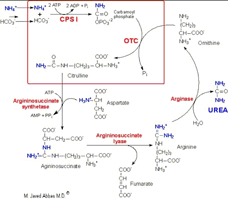
Select From The Following for More Details
Back to Topics<<<<

Introduction

Humans are totally dependent on other organisms for converting atmospheric nitrogen into forms available to the body. Nitrogen fixation is carried out by bacterial nitrogensases forming reduced nitrogen, NH4+ which can then be used by all organisms to form amino acids.

Overview of the flow of nitrogen in the biosphere. Nitrogen, nitrites and nitrates are acted upon by bacteria (nitrogen fixation) and plants and we assimilate these compounds as protein in our diets. Ammonia incorporation in animals occurs through the actions of glutamate dehydrogenase and glutamine synthase. Glutamate plays the central role in mammalian nitrogen flow, serving as both a nitrogen donor and nitrogen acceptor.

Reduced nitrogen enters the human body as dietary free amino acids, protein, and the ammonia produced by intestinal tract bacteria. A pair of principal enzymes, glutamate dehydrogenase and glutamine synthatase, are found in all organisms and effect the conversion of ammonia into the amino acids glutamate and glutamine, respectively. Amino and amide groups from these 2 substances are freely transferred to other carbon skeletons by transamination and transamidation reactions.

Representative aminotransferase catalyzed reaction.

Aminotransferases exist for all amino acids except threonine and lysine. The most common compounds involved as a donor/acceptor pair in transamination reactions are glutamate and a-ketoglutarate (a-KG), which participate in reactions with many different aminotransferases. Serum aminotransferases such as serum glutamate-oxaloacetate-aminotransferase (SGOT) (also called aspartate aminotransferase, AST) and serum glutamate-pyruvate aminotransferase (SGPT) (also called alanine transaminase, ALT) have been used as clinical markers of tissue damage, with increasing serum levels indicating an increased extent of damage. Alanine transaminase has an important function in the delivery of skeletal muscle carbon and nitrogen (in the form of alanine) to the liver. In skeletal muscle, pyruvate is transaminated to alanine, thus affording an additional route of nitrogen transport from muscle to liver. In the liver alanine transaminase tranfers the ammonia to a-KG and regenerates pyruvate. The pyruvate can then be diverted into gluconeogenesis. This process is refered to as the glucose-alanine cycle.
back to the top

The Glutamate Dehydrogenase Reaction

The reaction catalyzed by glutamate dehydrogenase is:

The glutamate dehydrogenase utilizes both nicotinamide nucleotide cofactors; NAD+ in the direction of nitrogen liberation and NADP+ for nitrogen incorporation. In the forward reaction as shown above glutamate dehydrogenase is important in converting free ammonia and a-ketoglutarate (a-KG) to glutamate, forming one of the 20 amino acids required for protein synthesis. However, it should be recognized that the reverse reaction is a key anapleurotic process linking amino acid metabolism with TCA cycle activity. In the reverse reaction, glutamate dehydrogenase provides an oxidizable carbon source used for the production of energy as well as a reduced electron carrier, NADH. As expected for a branch point enzyme with an important link to energy metabolism, glutamate dehydrogenase is regulated by the cell energy charge. ATP and GTP are positive allosteric effectors of the formation of glutamate, whereas ADP and GDP are positive allosteric effectors of the reverse reaction. Thus, when the level of ATP is high, conversion of glutamate to a-KG and other TCA cycle intermediates is limited; when the cellular energy charge is low, glutamate is converted to ammonia and oxidizable TCA cycle intermediates. Glutamate is also a principal amino donor to other amino acids in subsequent transamination reactions. The multiple roles of glutamate in nitrogen balance make it a gateway between free ammonia and the amino groups of most amino acids.
back to the top

The Glutamine Synthase Reaction

The reaction catalyzed by glutamine synthetase is:

glutamate + NH4+ + ATP -------> glutamine + ADP + Pi + H+

The glutamine synthetase reaction is also important in several respects. First it produces glutamine, one of the 20 major amino acids. Second, in animals, glutamine is the major amino acid found in the circulatory system. Its role there is to carry ammonia to and from various tissues but principally from peripheral tissues to the kidney, where the amide nitrogen is hydrolyzed by the enzyme glutaminase (reaction below); this process regenerates glutamate and free ammonium ion, which is excreted in the urine.

glutamine + H2O -------> glutamate + NH3

Note that, in this function, ammonia arising in peripheral tissue is carried in a nonionizable form which has none of the neurotoxic or alkalosis-generating properties of free ammonia.
Liver contains both glutamine synthetase and glutaminase but the enzymes are localized in different cellular segments. This ensures that the liver is neither a net producer nor consumer of glutamine. The differences in cellular location of these two enzymes allows the liver to scavange ammonia that has not been incorporated into urea. The enzymes of the urea cycle are located in the same cells as those that contain glutaminase. The result of the differential distribution of these two hepatic enzymes makes it possible to control ammonia incorporation into either urea or glutamine, the latter leads to excretion of ammonia by the kidney.
When acidosis occurs the body will divert more glutamine from the liver to the kidney. This allows for the conservation of bicarbonate ion since the incorporation of ammonia into urea requires bicarbonate (see below). When glutamine enters the kidney, glutaminase releases one mole of ammonia generating glutamate and then glutamate dehydrogenase releases another mole of ammonia generating a-ketoglutarate. The ammonia will ionizes to ammonium ion (NH4+) which is excreted. The net effect is a reduction in the pH (see also Kidneys and Acid-Base Balance).
back to the top

Digestive Tract Nitrogen

While glutamine, glutamate, and the remaining nonessential amino acids can be made by animals, the majority of the amino acids found in human tissues necessarily come from dietary sources (about 400g of protein per day). Protein digestion begins in the stomach, where a proenzyme called pepsinogen is secreted, autocatalytically converted to Pepsin A, and used for the first step of proteolysis. However, most proteolysis takes place in the duodenum as a consequence of enzyme activities secreted by the pancreas. All of the serine proteases and the zinc peptidases of pancreatic secretions are produced in the form of their respective proenzymes. These proteases are both endopeptidase and exopeptidase, and their combined action in the intestine leads to the production of amino acids, dipeptides, and tripeptides, all of which are taken up by enterocytes of the mucosal wall.
A circuitous regulatory pathway leading to the secretion of proenzymes into the intestine is triggered by the appearance of food in the intestinal lumen. Special mucosal endocrine cells secret the peptide hormones cholecystokinin (CCK) and secretin into the circulatory system. Together, CCK and secretin cause contraction of the gall bladder and the exocrine secretion of a bicarbonate-rich, alkaline fluid, containing protease proenzymes from the pancreas into the intestine. A second, paracrine role of CCK is to stimulate adjacent intestinal cells to secrete enteropeptidase, a protease that cleaves trypsinogen to produce trypsin. Trypsin also activates trypsinogen as well as all the other proenzymes in the pancreatic secretion, producing the active proteases and peptidases that hydrolyze dietary polypeptides.
Subsequent to luminal hydrolysis, small peptides and amino acids are transferred through enterocytes to the portal circulation by diffusion, facilitated diffusion, or active transport. A number of Na+-dependent amino acid transport systems with overlapping amino acid specificity have been described. In these transport systems, Na+ and amino acids at high luminal concentrations are co-transported down their concentration gradient to the interior of the cell. The ATP-dependent Na+/K+ pump exchanges the accumulated Na+ for extracellular K+, reducing intracellular Na+ levels and maintaining the high extracellular Na+ concentration (high in the intestinal lumen, low in enterocytes) required to drive this transport process.
Transport mechanisms of this nature are ubiquitous in the body. Small peptides are accumulated by a proton (H+) driven transport process and hydrolyzed by intracellular peptidases. Amino acids in the circulatory system and in extracellular fluids are transported into cells of the body by at least 7 different ATP-requiring active transport systems with overlapping amino acid specificities.
Hartnup disorder is an autosomal recessive impairment of neutral amino acid transport affecting the kidney tubules and small intestine. It is believed that the defect lies in a specific system responsible for neutral amino acid transport across the brush-border membrane of renal and intestinal epithelium. The exact defect has not yet been characterized. The characteristic diagnostic feature of Hartnup disorder is a dramatic neutral hyperaminoaciduria. Additionally, individuals excrete indolic compounds that originate from the bacterial degradation of unabsorbed tryptophan. The reduced intestinal absorption and increased renal loss of tryptophan lead to a reduced availability of tryptophan for niacin and nicotinamide nucleotide biosynthesis. As a consequence affected individuals frequently exhibit pellegra-like rashes.
Many other nitrogenous compounds are found in the intestine. Most are bacterial products of protein degradation. Some have powerful pharmacological (vasopressor) effects.

Products of Intestinal Bacterial Activity
Substrates Products
Vasopressor Amines Other
Lysine Cadaverene  
Arginine Agmatine  
Tyrosine Tyramine  
Ornithine Putrescine  
Histidine Histamine  
Tryptophan   Indole and skatole
All amino acids   NH4+

Prokaryotes such as E. coli can make the carbon skeletons of all 20 amino acids and transaminate those carbon skeletons with nitrogen from glutamine or glutamate to complete the amino acid structures. Humans cannot synthesize the branched carbon chains found in branched chain amino acids or the ring systems found in phenylalanine and the aromatic amino acids; nor can we incorporate sulfur into covalently bonded structures. Therefore, the 10 so-called essential amino acids (see Table below) must be supplied from the diet. Nevertheless, it should be recognized that, depending on the composition of the diet and physiological state of an individual, one or another of the non-essential amino acids may also become a required dietary component. For example, arginine is only normally considered to be essential amino acid during early childhood development because enough for adult needs is made by the urea cycle.
To take a different type of example, cysteine and tyrosine are considered non-essential but are formed from the essential amino acids methionine and phenylalanine, respectively. If sufficient cysteine and tyrosine are present in the diet, the requirements for methionine and phenylalanine are markedly reduced; conversely, if methionine and phenylalanine are present in only limited quantities, cysteine and tyrosine can become essential dietary components. Finally, it should be recognized that if the a-keto acids corresponding to the carbon skeletons of the essential amino acids are supplied in the diet, aminotransferases in the body will convert the keto acids to their respective amino acids, largely supplying the basic needs.
Unlike fats and carbohydrates, nitrogen has no designated storage depots in the body. Since the half-life of many proteins is short (on the order of hours), insufficient dietary quantities of even one amino acid can quickly limit the synthesis and lower the body levels of many essential proteins. The result of limited synthesis and normal rates of protein degradation is that the balance of nitrogen intake and nitrogen excretion is rapidly and significantly altered. Normal, healthy adults are generally in nitrogen balance, with intake and excretion being very well matched. Young growing children, adults recovering from major illness, and pregnant women are often in positive nitrogen balance. Their intake of nitrogen exceeds their loss as net protein synthesis proceeds. When more nitrogen is excreted than is incorporated into the body, an individual is in negative nitrogen balance. Insufficient quantities of even one essential amino acid is adequate to turn an otherwise normal individual into one with a negative nitrogen balance.
The biological value of dietary proteins is related to the extent to which they provide all the necessary amino acids. Proteins of animal origin generally have a high biological value; plant proteins have a wide range of values from almost none to quite high. In general, plant proteins are deficient in lysine, methionine, and tryptophan and are much less concentrated and less digestible than animal proteins. The absence of lysine in low-grade cereal proteins, used as a dietary mainstay in many underdeveloped countries, leads to an inability to synthesize protein (because of missing essential amino acids) and ultimately to a syndrome known as kwashiorkor, common among children in these countries.
back to the top

Essential vs. Nonessential Amino Acids

Nonessential Essential
Alanine Arginine*
Asparagine Histidine
Aspartate Isoleucine
Cysteine Leucine
Glutamate Lysine
Glutamine Methionine*
Glycine Phenylalanine*
Proline Threonine
Serine Tyrptophan
Tyrosine Valine

*The amino acids arginine, methionine and phenylalanine are considered essential for reasons not directly related to lack of synthesis. Arginine is synthesized by mammalian cells but at a rate that is insufficient to meet the growth needs of the body and the majority that is synthesized is cleaved to form urea. Methionine is required in large amounts to produce cysteine if the latter amino acid is not adequately supplied in the diet. Similarly, phenyalanine is needed in large amounts to form tyrosine if the latter is not adequately supplied in the diet.

back to the top

Removal of Nitrogen from Amino Acids

The dominant reactions involved in removing amino acid nitrogen from the body are known as transaminations. This class of reactions funnels nitrogen from all free amino acids into a small number of compounds; then, either they are oxidatively deaminated, producing ammonia, or their amine groups are converted to urea by the urea cycle. Transaminations involve moving an a-amino group from a donor a-amino acid to the keto carbon of an acceptor a-keto acid. These reversible reactions are catalyzed by a group of intracellular enzymes known as aminotransferases, which generally employ covalently bound pyridoxal phosphate as a cofactor. However, some aminotransferases employ pyruvate as a cofactor.
Aminotransferases exist for all amino acids except threonine and lysine. The most common compounds involved as a donor/acceptor pair in transamination reactions are glutamate and a-ketoglutarate, which participate in reactions with many different aminotransferases. Serum aminotransferases such as serum glutamate-oxaloacetate-aminotransferase (SGOT) (also called aspartate aminotransferase, AST) and serum glutamate-pyruvate aminotransferase (SGPT) (also called alanine transaminase, ALT) have been used as clinical markers of tissue damage, with increasing serum levels indicating an increased extent of damage. Alanine transaminase has an important function in the delivery of skeletal muscle carbon and nitrogen (in the form of alanine) to the liver. In skeletal muscle, pyruvate is transaminated to alanine, thus affording an additional route of nitrogen transport from muscle to liver. In the liver alanine transaminase tranfers the ammonia to a-KG and regenerates pyruvate. The pyruvate can then be diverted into gluconeogenesis. This process is refered to as the glucose-alanine cycle.
Because of the participation of a-ketoglutarate in numerous transaminations, glutamate is a prominent intermediate in nitrogen elimination as well as in anabolic pathways. Glutamate formed in the course of nitrogen elimination is either oxidatively deaminated by liver glutamate dehydrogenase, forming ammonia, or converted to glutamine by glutamine synthase and transported to kidney tubule cells. There the glutamine is sequentially deamidated by glutaminase and deaminated by kidney glutamate dehydrogenase.
The ammonia produced in the latter two reactions is excreted as NH4+ in the urine, where it helps maintain urine pH in the normal range of pH 4 to pH 8. The extensive production of ammonia by peripheral tissue or hepatic glutamate dehydrogenase is not feasible because of the highly toxic effects of circulating ammonia. Normal serum ammonium concentrations are in the range of 20 - 40 mM, and an increase in circulating ammonia to about 400 mM causes alkalosis and neurotoxicity.
A final, therapeutically useful amino acid-related reaction is the amidation of aspartic acid to produce asparagine. The enzyme asparagine synthase catalyzes the ATP-requiring transamidation reaction shown below:

aspartate + glutamine + ATP -----> glutamate + asparagine + AMP + PPi

Most cells perform this reaction well enough to produce all the asparagine they need. However, some leukemia cells require exogenous asparagine, which they obtain from the plasma. Chemotherapy using the enzyme asparaginase takes advantage of this property of leukemic cells by hydrolyzing serum asparagine to ammonia and aspartic acid, thus depriving the neoplastic cells of the asparagine that is essential for their characteristic rapid growth.
In the peroxisomes of mammalian tissues, especially liver, there exist a minor enzymatic pathway for the removal of amino groups from amino acids. L-amino acid oxidase is FMN-linked and has broad specificity for the L-amino acids.
A number of substances, including oxygen, can act as electron acceptors from the flavoproteins. If oxygen is the acceptor the product is hydrogen peroxide, which is then rapidly degraded by the catalases found in liver and other tissues.
Missing or defective biogenesis of peroxisomes or L-amino acid oxidase causes generalized hyperaminoacidemia and hyperaminoaciduria, generally leading to neurotoxicity and early death.
back to the top

The Urea Cycle

Earlier it was noted that kidney glutaminase was responsible for converting excess glutamine from the liver to urine ammonium. However, about 80% of the excreted nitrogen is in the form of urea which is also largely made in the liver, in a series of reactions that are distributed between the mitochondrial matrix and the cytosol. The series of reactions that form urea is known as the Urea Cycle or the Krebs-Henseleit Cycle.

Diagram of the urea cycle. The reactions of the urea cycle which occur in the mitochondrion are contained in the red rectangle. All enzymes are in red, CPS-I is carbamoyl phosphate synthetase-I, OTC is ornithine transcarbamoylase.
The essential features of the urea cycle reactions and their metabolic regulation are as follows: Arginine from the diet or from protein breakdown is cleaved by the cytosolic enzyme arginase, generating urea and ornithine. In subsequent reactions of the urea cycle a new urea residue is built on the ornithine, regenerating arginine and perpetuating the cycle.
Ornithine arising in the cytosol is transported to the mitochondrial matrix, where ornithine transcabamoylase catalyzes the condensation of ornithine with carbamoyl phosphate, producing citrulline. The energy for the reaction is provided by the high-energy anhydride of carbamoyl phosphate. The product, citrulline, is then transported to the cytosol, where the remaining reactions of the cycle take place.
The synthesis of citrulline requires a prior activation of carbon and nitrogen as carbamoyl phosphate (CP). The activation step requires 2 equivalents of ATP and the mitochondrial matrix enzyme carbamoyl phosphate synthetase-I (CPS-I).There are two CP synthetases: a mitochondrial enzyme, CPS-I, which forms CP destined for inclusion in the urea cycle, and a cytosolic CP synthatase (CPS-II), which is involved in pyrimidine nucleotide biosynthesis. CPS-I is positively regulated by the allosteric effector N-acetyl-glutamate, while the cytosolic enzyme is acetylglutamate independent.
In a 2-step reaction, catalyzed by cytosolic argininosuccinate synthetase, citrulline and aspartate are condensed to form argininosuccinate. The reaction involves the addition of AMP (from ATP) to the amido carbonyl of citrulline, forming an activated intermediate on the enzyme surface (AMP-citrulline), and the subsequent addition of aspartate to form argininosuccinate.
Arginine and fumarate are produced from argininosuccinate by the cytosolic enzyme argininosuccinate lyase (also called argininosuccinase). In the final step of the cycle arginase cleaves urea from aspartate, regenerating cytosolic ornithine, which can be transported to the mitochondrial matrix for another round of urea synthesis. The fumarate, generated via the action of arginiosuccinate lyase, is reconverted to aspartate for use in the argininosuccinate synthetase reaction. This occurs through the actions of cytosolic versions of the TCA cycle enzymes, fumarase (which yields malate) and malate dehydrogenase (which yields oxaloacetate). The oxaloacetate is then transaminated to aspartate by AST.
Beginning and ending with ornithine, the reactions of the cycle consumes 3 equivalents of ATP and a total of 4 high-energy nucleotide phosphates. Urea is the only new compound generated by the cycle; all other intermediates and reactants are recycled. The energy consumed in the production of urea is more than recovered by the release of energy formed during the synthesis of the urea cycle intermediates. Ammonia released during the glutamate dehydrogenase reaction is coupled to the formation of NADH. In addition, when fumarate is converted back to aspartate, the malate dehydrogenase reaction used to convert malate to oxaloacetate generates a mole of NADH. These two moles of NADH, thus, are oxidized in the mitochondria yielding 6 moles of ATP.
back to the top

Regulation of the Urea Cycle

The urea cycle operates only to eliminate excess nitrogen. On high-protein diets the carbon skeletons of the amino acids are oxidized for energy or stored as fat and glycogen, but the amino nitrogen must be excreted. To facilitate this process, enzymes of the urea cycle are controlled at the gene level. With long-term changes in the quantity of dietary protein, changes of 20-fold or greater in the concentration of cycle enzymes are observed. When dietary proteins increase significantly, enzyme concentrations rise. On return to a balanced diet, enzyme levels decline. Under conditions of starvation, enzyme levels rise as proteins are degraded and amino acid carbon skeletons are used to provide energy, thus increasing the quantity of nitrogen that must be excreted.
Short-term regulation of the cycle occurs principally at CPS-I, which is relatively inactive in the absence of its allosteric activator N-acetylglutamate. The steady-state concentration of N-acetylglutamate is set by the concentration of its components acetyl-CoA and glutamate and by arginine, which is a positive allosteric effector of N-acetylglutamate synthetase.

acetyl-CoA + glutamate ----> N-acetylglutamate + CoA


back to the top


Urea Cycle Defects (UCDs)

A complete lack of any one of the enzymes of the urea cycle will result in death shortly after birth. However, deficiencies in each of the enzymes of the urea cycle, including N-acetylglutamate synthase, have been identified. These disorders are referred to as urea cycle disorders or UCDs. More information on the individual UCDs can be found in the Inborn Errors in Metabolism pages. A common thread to most UCDs is hyperammonemia leading to ammonia intoxication with the consequences described below. Deficiencies in arginase do not lead to symptomatic hyperammonemia as severe or as commonly as in the other UCDs.
Clinical symptoms are most severe when the UCD is at the level of carbamoyl phosphate synthetase I. Symptoms of UCDs usually arise at birth and encompass, ataxia, convulsions, lethargy, poor feeding and eventually coma and death if not recognized and treated properly. In fact, the mortality rate is 100% for UCDs that are left undiagnosed. Several UCDs manifest with late-onset such as in adulthood. In these cases the symptoms are hyperactivity, hepatomegaly and an avoidance of high protein foods.
In general, the treatment of UCDs has as common elements the reduction of protein in the diet, removal of excess ammonia and replacement of intermediates missing from the urea cycle. Administration of levulose reduces ammonia through its action of acidifying the colon. Bacteria metabolize levulose to acidic byproducts which then promotes excretion of ammonia in the feces as ammonium ions, NH4+. Antibiotics can be administered to kill intestinal ammonia producing bacteria. Sodium benzoate and sodium phenylbutyrate can be administered to covalently bind glycine (forming hippurate) and glutamine (forming phenylacetylglutamine), respectively. These latter compounds, which contain the ammonia nitrogen, are excreted in the feces. Dietary supplementation with arginine or citrulline can increase the rate of urea production in certain UCDs.
back to the top

Table of UCDs

UCDEnzyme DeficiencySymptoms/Comments
Type I HyperammonemiaCarbamoylphosphate synthetase Iwith 24h - 72h after birth infant becomes lethargic, needs stimulation to feed, vomiting, increasing lethargy, hypothermia and hyperventilation; without measurement of serum ammonia levels and appropriate intervention infant will die: treament with arginine which activates N-acetylglutamate synthetase
N-acetylglutamate synthetase DeficiencyN-acetylglutamate synthetasesevere hyperammonemia, mild hyperammonemia associated with deep coma, acidosis, recurrent diarrhea, ataxia, hypoglycemia, hyperornithinemia: treatment includes administration of carbamoyl glutamate to activate CPS I
Type 2 HyperammonemiaOrnithine transcarbamoylasemost commonly occurring UCD, only X-linked UCD, ammonia and amino acids elevated in serum, increased serum orotic acid due to mitochondrial carbamoylphosphate entering cytosol and being incorporated into pyrimidine nucleotides which leads to excess production and consequently excess catabolic products: treat with high carbohydrate, low protein diet, ammonia detoxification with sodium phenylacetate or sodium benzoate
Classic CitrullinemiaArgininosuccinate synthetaseepisodic hyperammonemia, vomiting, lethargy, ataxia, siezures, eventual coma: treat with arginine administration to enhance citrulline excretion, also with sodium benzoate for ammonia detoxification
Argininosuccinic aciduriaArgininosuccinate lyase
(argininosuccinase)
episodic symptoms similar to classic citrullinemia, elevated plasma and cerebral spinal fluid argininosuccinate: treat with arginine and sodium benzoate
HyperargininemiaArginaserare UCD, progressive spastic quadriplegia and mental retardation, ammonia and arginine high in cerebral spinal fluid and serum, arginine, lysine and ornithine high in urine: treatment includes diet of essential amino acids excluding arginine, low protein diet

back to the top

Neurotoxicity Associated with Ammonia

Earlier it was noted that ammonia was neurotoxic. Marked brain damage is seen in cases of failure to make urea via the urea cycle or to eliminate urea through the kidneys. The result of either of these events is a buildup of circulating levels of ammonium ion. Aside from its effect on blood pH, ammonia readily traverses the brain blood barrier and in the brain is converted to glutamate via glutamate dehydrogenase, depleting the brain of a-ketoglutarate. As the a-ketoglutarate is depleted, oxaloacetate falls correspondingly, and ultimately TCA cycle activity comes to a halt. In the absence of aerobic oxidative phosphorylation and TCA cycle activity, irreparable cell damage and neural cell death ensue. In addition, the increased glutamate leads to glutamine formation. This depletes glutamate stores which are needed in neural tissue since glutamate is both a neurotransmitter and a precursor for the synthesis of g-aminobutyrate, GABA, another neurotransmitter. Therefore, reductions in brain glutamate affect energy production as well as neurotransmission.
back to the top
Back to Topics<<<<


This article has been modified by Dr. M. Javed Abbas.
If you have any comments please do not hesitate to sign my Guest Book.

20:47 21/12/2002