Select From The Following for More Details
Back to Topics<<<<

The Pyruvate Dehydrogenase (PDH) Complex

The bulk of ATP used by many cells to maintain homeostasis is produced by the oxidation of pyruvate in the TCA cycle. During this oxidation process, reduced nicotinamide adenine dinucleotide (NADH) and reduced flavin adenine dinucleotide (FADH2) are generated. The NADH and FADH2 are principally used to drive the processes of oxidative phosphorylation, which are responsible for converting the reducing potential of NADH and FADH2 to the high energy phosphate in ATP.
The fate of pyruvate depends on the cell energy charge. In cells or tissues with a high energy charge pyruvate is directed toward gluconeogenesis, but when the energy charge is low pyruvate is preferentially oxidized to CO2 and H2O in the TCA cycle, with generation of 15 equivalents of ATP per pyruvate. The enzymatic activities of the TCA cycle (and of oxidative phosphorylation) are located in the mitochondrion. When transported into the mitochondrion, pyruvate encounters two principal metabolizing enzymes: pyruvate carboxylase (a gluconeogenic enzyme) and pyruvate dehydrogenase (PDH), the first enzyme of the PDH complex. With a high cell-energy charge coenzyme A (CoA) is highly acylated, principally as acetyl-CoA, and able allosterically to activate pyruvate carboxylase, directing pyruvate toward gluconeogenesis. When the energy charge is low CoA is not acylated, pyruvate carboxylase is inactive, and pyruvate is preferentially metabolized via the PDH complex and the enzymes of the TCA cycle to CO2 and H2O. Reduced NADH and FADH2 generated during the oxidative reactions can then be used to drive ATP synthesis via oxidative phosphorylation.
The PDH complex is comprised of multiple copies of 3 separate enzymes: pyruvate dehydrogenase (20-30 copies), dihydrolipoyl transacetylase (60 copies) and dihydrolipoyl dehydrogenase (6 copies). The complex also requires 5 different coenzymes: CoA, NAD+, FAD+, lipoic acid and thiamine pyrophosphate (TPP) . Three of the coenzymes of the complex are tightly bound to enzymes of the complex (TPP, lipoic acid and FAD+) and two are employed as carriers of the products of PDH complex activity (CoA and NAD+). The pathway of PDH oxidation of pyruvate to acetyl-CoA is diagrammed below.

Flow diagram depicting the overall activity of the pyruvate dehydrogenase complex. During the oxidation of pyruvate to CO2 by pyruvate dehydrogenase the electrons flow from pyruvate to the lipoamide moiety of dihydrolipoyl transacetylase then to the FAD cofactor of dihydrolipoyl dehydrogenase and finally to reduction of NAD+ to NADH. The acetyl group is linked to coenzyme A (CoASH) in a high energy thioester bond. The acetyl-CoA then enters the TCA cycle for complete oxidation to CO2 and H2O.
The first enzyme of the complex is PDH itself which oxidatively decarboxylates pyruvate. During the course of the reaction the acetyl group derived from decarboxylation of pyruvate is bound to TPP. The next reaction of the complex is the transfer of the 2--carbon acetyl group from acetyl-TPP to lipoic acid, the covalently bound coenzyme of lipoyl transacetylase. The transfer of the acetyl group from acyl-lipoamide to CoA results in the formation of 2 sulfhydryl (SH) groups in lipoate requiring reoxidation to the disulfide (S-S) form to regenerate lipoate as a competent acyl acceptor. The enzyme dihydrolipoyl dehydrogenase, with FAD+ as a cofactor, catalyzes that oxidation reaction. The final activity of the PDH complex is the transfer of reducing equivalents from the FADH2 of dihydrolipoyl dehydrogenase to NAD+. The fate of the NADH is oxidation via mitochondrial electron transport, to produce 3 equivalents of ATP:
The net result of the reactions of the PDH complex are:

Pyruvate + CoA + NAD+ ------> CO2 + acetyl-CoA + NADH + H+

back to the top


Regulation of the PDH Complex

The reactions of the PDH complex serves to interconnect the metabolic pathways of glycolysis, gluconeogenesis and fatty acid synthesis to the TCA cycle. As a consequence, the activity of the PDH complex is highly regulated by a variety of allosteric effectors and by covalent modification. The importance of the PDH complex to the maintenance of homeostasis is evident from the fact that although diseases associated with deficiencies of the PDH complex have been observed, affected individuals often do not survive to maturity. Since the energy metabolism of highly aerobic tissues such as the brain is dependent on normal conversion of pyruvate to acetyl-CoA, aerobic tissues are most sensitive to deficiencies in components of the PDH complex. Most genetic diseases associated with PDH complex deficiency are due to mutations in PDH. The main pathologic result of such mutations is moderate to severe cerebral lactic acidosis and encephalopathies.
The main regulatory features of the PDH complex are diagrammed below.
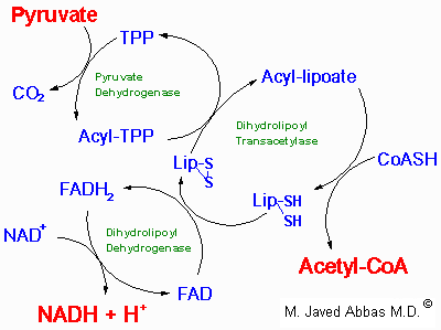
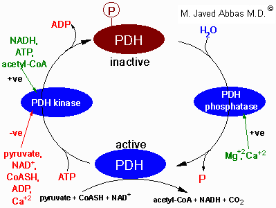
Factors regulating the activity of pyruvate dehydrogenase, (PDH). PDH activity is regulated by its' state of phosphorylation, being most active in the dephosphorylated state. Phosphorylation of PDH is catalyzed by a specific PDH kinase. The activity of the kinase is enhanced when cellular energy charge is high which is reflected by an increase in the level of ATP, NADH and acetyl-CoA. Conversely, an increase in pyruvate strongly inhibits PDH kinase. Additional negative effectors of PDH kinase are ADP, NAD+ and CoASH, the levels of which increase when energy levels fall. The regulation of PDH phosphatase is not completely understood but it is known that Mg2+ and Ca2+ activate the enzyme. In adipose tissue insulin increases PDH activity and in cardiac muscle PDH activity is increased by catecholamines.
Two products of the complex, NADH and acetyl-CoA, are negative allosteric effectors on PDH-a, the non-phosphorylated, active form of PDH. These effectors reduce the affinity of the enzyme for pyruvate, thus limiting the flow of carbon through the PDH complex. In addition, NADH and acetyl-CoA are powerful positive effectors on PDH kinase, the enzyme that inactivates PDH by converting it to the phosphorylated PDH-b form. Since NADH and acetyl-CoA accumulate when the cell energy charge is high, it is not surprising that high ATP levels also up-regulate PDH kinase activity, reinforcing down-regulation of PDH activity in energy-rich cells. Note, however, that pyruvate is a potent negative effector on PDH kinase, with the result that when pyruvate levels rise, PDH-a will be favored even with high levels of NADH and acetyl-CoA.
Concentrations of pyruvate which maintain PDH in the active form (PDH-a) are sufficiently high so that, in energy-rich cells, the allosterically down-regulated, high Km form of PDH is nonetheless capable of converting pyruvate to acetyl-CoA. With large amounts of pyruvate in cells having high energy charge and high NADH, pyruvate carbon will be directed to the 2 main storage forms of carbon---glycogen via gluconeogenesis and fat production via fatty acid synthesis---where acetyl-CoA is the principal carbon donor.
Although the regulation of PDH-b phosphatase is not well understood, it is quite likely regulated to maximize pyruvate oxidation under energy-poor conditions and to minimize PDH activity under energy-rich conditions.
back to the top

Reactions of the TCA Cycle

The TCA cycle showing enzymes, substrates and products. The abbreviated enzymes are: IDH = isocitrate dehydrogenase and a-KGDH = a-ketoglutarate dehydrogenase. The GTP generated during the succinate thiokinase (succinyl-CoA synthetase) reaction is equivalent to a mole of ATP by virtue of the presence of nucleoside diphosphokinase. The 3 moles of NADH and 1 mole of FADH2 generated during each round of the cycle feed into the oxidative phosphorylation pathway. Each mole of NADH leads to 3 moles of ATP and each mole of FADH2 leads to 2 moles of ATP. Therefore, for each mole of pyruvate which enters the TCA cycle, 12 moles of ATP can be generated.

Citrate Synthase (Condensing enzyme)

The first reaction of the cycle is condensation of the methyl carbon of acetyl-CoA with the keto carbon (C-2) of oxaloacetate (OAA). The standard free energy of the reaction, -8.0 kcal/mol, drives it strongly in the forward direction. Since the formation of OAA from its precursor is thermodynamically unfavorable, the highly exergonic nature of the citrate synthase reaction is of central importance in keeping the entire cycle going in the forward direction, since it drives oxaloacetate formation by mass action principals.
When the cellular energy charge increases the rate of flux through the TCA cycle will decline leading to a build-up of citrate. Excess citrate is used to transport acetyl-CoA carbons from the mitochondrion to the cytoplasm where they can be used for fatty acid and cholesterol biosynthesis. Additionally, the increased levels of citrate in the cytoplasm activate the key regulatory enzyme of fatty acid biosynthesis, acetyl-CoA carboxylase (ACC) and inhibit PFK-1. In non-hepatic tissues citrate is also required for ketone body synthesis.

Aconitase

The isomerization of citrate to isocitrate by aconitase is stereospecific, with the migration of the -OH from the central carbon of citrate (formerly the keto carbon of OAA) being always to the adjacent carbon which is derived from the methylene (-CH2-) of OAA. The stereospecific nature of the isomerization determines that the CO2 lost, as isocitrate is oxidized to succinyl-CoA, is derived from the oxaloacetate used in citrate synthesis.
Aconitase is one of several mitochondrial enzymes known as non-heme-iron proteins. These proteins contain inorganic iron and sulfur, known as iron sulfur centers, in a coordination complex with cysteine sulfurs of the protein. There are two prominent classes of non-heme-iron complexes, those containing two equivalents each of inorganic iron and sulfur Fe2S2, and those containing 4 equivalents of each Fe4S4. Aconitase is a member of the Fe4S4 class. Its iron sulfur centers are often designated as Fe4S4Cys4, indicating that 4 cystine sulfur atoms are involved in tghe complete structure of the complex. In iron sulfur compounds the iron is generally involved in oxidation-reduction events.

Isocitrate Dehydrogenase

Isocitrate is oxidatively decarboxylated to a-ketoglutarate by isocitrate dehydrogenase, (IDH). There are two different IDH enzymes. The IDH of the TCA cycle uses NAD+ as a cofactor, whereas the other IDH uses NADP+ as a cofactor. Unlike the NAD+-requiring enzyme, which is located only in the mitochondrial matrix, the NADP+-requiring enzyme is found in both the mitochondrial matrix and the cytosol. IDH catalyzes the rate-limiting step, as well as the first NADH-yielding reaction of the TCA cycle. The CO2 produced by the IDH reaction is the original C-1 of the oxaloacetate used in the citrate synthase reaction.
It is generally considered that control of carbon flow through the cycle is regulated at IDH by the powerful negative allosteric effectors NADH and ATP and by the potent positive effectors; isocitrate, ADP and AMP. From the latter it is clear that cell energy charge is a key factor in regulating carbon flow through the TCA cycle.

a-Ketoglutarate Dehydrogenase Complex

a-ketoglutarate is oxidatively decarboxylated to succinyl-CoA by the a-ketoglutarate dehydrogenase (a-KGDH) complex. This reaction generates the second TCA cycle equivalent of CO2 and NADH. This multienzyme complex is very similar to the PDH complex in the intricacy of its protein makeup, cofactors, and its mechanism of action. Also, as with the PDH complex, the reactions of the a-KGDH complex proceed with a large negative standard free energy change. Although the a-KGDH of the complex is not subject to covalent modification, allosteric regulation is quite complex, with activity being regulated by energy charge, the NAD+/NADH ratio, and effector activity of substrates and products.
Succinyl-CoA and a-ketoglutarate are also important metabolites outside the TCA cycle. In particular, a-ketoglutarate represents a key anapleurotic metabolite linking the entry and exit of carbon atoms from the TCA cycle to pathways involved in amino acid metabolism. a-ketoglutarate is also important for driving the malate-aspartate shuttle. Succinyl-CoA, along with glycine, contributes all the carbon and nitrogen atoms required for the synthesis of protoporphyrin heme biosynthesis and for non-hepatic tissue utilization of ketone bodies.

Succinyl CoA Synthetase (Succinyl Thiokinase )

The conversion of succinyl-CoA to succinate by succinyl CoA synthetase involves use of the high-energy thioester of succinyl-CoA to drive synthesis of a high-energy nucleotide phosphate, by a process known as substrate-level phosphorylation. In this process a high energy enzyme--phosphate intermediate is formed, with the phosphate subsequently being transferred to GDP. Mitochondrial GTP is used in a trans-phosphorylation reaction catalyzed by the mitochondrial enzyme nucleoside diphospho kinase to phosphorylate ADP, producing ATP and regenerating GDP for the continued operation of succinyl CoA synthetase.

Succinate Dehydrogenase (SDH)

Succinate dehydrogenase catalyzes the oxidation of succinate to fumarate with the sequential reduction of enzyme-bound FAD and non-heme-iron. In mammalian cells the final electron acceptor is coenzyme Q10 (CoQ10), a mobile carrier of reducing equivalents that is restricted by its lipophilic nature to the lipid phase of the mitochondrial membrane.

Fumarase (fumarate hydratase)

The fumarase-catalyzed reactions specific for the trans form of fumarate. The result is that the hydration of fumarate proceeds stereospecifically with the production of L-malate.

Malate Dehydrogenase (MDH)

L-malate is the specific substrate for MDH, the final enzyme of the TCA cycle. The forward reaction of the cycle, the oxidation of malate yields oxaloacetate (OAA). In the forward direction the reaction has a standard free energy of about +7 kcal/mol, indicating the very unfavorable nature of the forward direction. As noted earlier, the citrate synthase reaction that condenses oxaloacetate with acetyl-CoA has a standard free energy of about -8 kcal/mol and is responsible for pulling the MDH reaction in the forward direction. The overall change in standard free energy change is about -1 kcal/mol for the conversion of malate to oxaloacetate and on to succinate.
The overall stoichiometry of the TCA cycle is:

acetyl-CoA + 3NAD+ + FAD + GDP + Pi + 2H2O ----> 2CO2 + 3NADH + FADH2 + GTP + 2H+ + HSCoA

back to the top


Regulation of the TCA Cycle

Regulation of the TCA cycle. like that of glycolysis, occurs at both the level of entry of substrates into the cycle as well as at the key reactions of the cycle. Fuel enters the TCA cycle primarily as acetyl-CoA. The generation of acetyl-CoA from carbohydrates is, therefore, a major control point of the cycle. This is the reaction catalyzed by the PDH complex.
By way of review, the PDH complex is inhibited by acetyl-CoA and NADH and activated by non-acetylated CoA (CoASH) and NAD+. The pyruvate dehydrogenase activities of the PDH complex are regulated by their state of phosphorylation. This modification is carried out by a specific kinase (PDH kinase) and the phosphates are removed by a specific phosphatase (PDH phosphatase). The phosphorylation of PDH inhibits its activity and, therefore, leads to decreased oxidation of pyruvate. PDH kinase is activated by NADH and acetyl-CoA and inhibited by pyruvate, ADP, CoASH, Ca2+ and Mg2+. The PDH phosphatase, in contrast, is activated by Mg2+ and Ca2+.
Since three reactions of the TCA cycle as well as PDH utilize NAD+ as co-factor it is not difficult to understand why the cellular ratio of NAD+/NADH has a major impact on the flux of carbon through the TCA cycle.
Substrate availability can also regulate TCA flux. This occurs at the citrate synthase reaction as a result of reduced availability of oxaloacetate. Product inhibition also controls the TCA flux, e.g. citrate inhibits citrate synthase, a-KGDH is inhibited by NADH and succinyl-CoA. The key enzymes of the TCA cycle are also regulated allosterically by Ca2+, ATP and ADP.
back to the top
Back to Topics<<<<


This article has been modified by Dr. M. Javed Abbas.
If you have any comments please do not hesitate to sign my Guest Book.

20:40 21/12/2002