Select From The Following for More Details
Back to Topics<<<<

Introduction
Translation is the RNA directed synthesis of polypeptides. This process requires all three classes of RNA. Although the chemistry of peptide bond formation is relatively simple, the processes leading to the ability to form a peptide bond are exceedingly complex. The template for correct addition of individual amino acids is the mRNA, yet both tRNAs and rRNAs are involved in the process. The tRNAs carry activated amino acids into the ribosome which is composed of rRNA and ribosomal proteins. The ribosome is associated with the mRNA ensuring correct access of activated tRNAs and containing the necessary enzymatic activities to catalyze peptide bond formation.
back to the top
Historical Perspectives
Early genetic experiments demonstrated:
  • 1. the colinearity between the DNA and protein encoded by the DNA. Yanofsky showed that order of observed mutations in the E. coli tryptophan synthetase gene was the same as the corresponding amino acid changes in the protein.
  • 2. Crick and Brenner demonstrated, from a large series of double mutants of the bacteriophage T4, that the genetic code is read in a sequential manner starting from a fixed point in the gene, the code was most likely a triplet and that all 64 possible combinations of the 4 nucleotides code for amino acids, i.e. the code is degenerate since there are only 20 amino acids.
The above mentioned experiments only indicated deductive correlation's regarding the genetic code. The precise dictionary of the genetic code was originally determined by the use of in vitro translation systems derived from E. coli cells. Synthetic polyribonucleotides were added to these translation system along with all twenty amino acids. One amino acid at a time was radiolabeled. The first demonstration of the dictionary of the genetic code was with the use of poly(U). This synthetic polyribonucleotide encoded the amino acid phenylalanine, i.e. the resulting polypeptide was poly(F).
The utilization of a variety of repeating di- tri- and tetra polyribonucleotides established the entire genetic code. These results of these experiments confirmed that some amino acids are encoded for by more than one triplet codon, hence the degeneracy of the genetic code. These experiments also established the identity of translational termination codons.
An additional important point to come from these early experiments was that the 5' end of the RNA corresponded to the amino terminus of the polypeptide. This was important since previous labeling experiments had demonstrated that the N-terminus is the beginning of the elongating polypeptide. Therefore, in vitro translation experiments established that the RNA is read in the 5' to 3' direction.
Crick first postulated that translation of the genetic code would be carried out through mediation of adapter molecules. Each adapter was postulated to carry a specific amino acid and to recognize the corresponding codon. He suggested that the adapters contain RNA because codon recognition could then occur by complimentarity to the sequences of the codons in the mRNA.
During the course of in vitro protein synthesis and labeling experiments it was shown that the amino acids became transiently bound to a low molecular weight mass fraction of RNA. This fraction of RNAs have been termed transfer RNAs (tRNAs) since they transfer amino acids to the elongating polypeptide. These results indicate that accurate translation requires two equally important recognition steps:

  • 1. the correct choice of amino acid needs to be made for attachment to the correspondingly correct tRNA.
  • 2. selection of the correct amino acid-charged tRNA by the mRNA. This process is facilitated by the ribosomes which we will discuss below.

Summary of Experiments to Determine the Genetic Code

  • 1. The genetic code is read in a sequential manner starting near the 5' end of the mRNA. This means that translation proceeds along the mRNA in the 5' ---> 3' direction which corresponds to the N-terminal to C-terminal direction of the amino acid sequences within proteins.
  • 2. The code is composed of a triplet of nucleotides.
  • 3. That all 64 possible combinations of the 4 nucleotides code for amino acids, i.e. the code is degenerate since there are only 20 amino acids.

The precise dictionary of the genetic code was determined with the use of in vitro translation systems and polyribonucleotides. The results of these experiments confirmed that some amino acids are encoded by more than one triplet codon, hence the degeneracy of the genetic code. These experiments also established the identity of translational termination codons.
back to the top
The Genetic Code
U C A G
  U UUU Phe
UU
C Phe
UU
A Leu
UU
G Leu
UCU Ser
UC
C Ser
UC
A Ser
UC
G Ser
UAU Tyr
UA
C Tyr
UAA End
UAG End
UGU Cys
UG
C Cys
UGA End
UGG Trp
 
C
CUU Leu
CU
C Leu
CU
A Leu
CU
G Leu
CCU Pro
CC
C Pro
CC
A Pro
CC
G Pro
CAU His
CA
C His
CA
A Gln
CA
G Gln
CGU Arg
CG
C Arg
CG
A Arg
CG
G Arg
 
A
AUU Ile
AU
C Ile
AU
A Ile
AU
G Met
ACU Thr
AC
C Thr
AC
A Thr
AC
G Thr
AAU Asn
AA
C Asn
AA
A Lys
AA
G Lys
AGU Ser
AG
C Ser
AG
A Arg
AG
G Arg
 
G
GUU Val
GU
C Val
GU
A Val
GU
G Val
GCU Ala
GC
C Ala
GC
A Ala
GC
G Ala
GAU Asp
GA
C Asp
GA
A Glu
GA
G Glu
GGU Gly
GG
C Gly
GG
A Gly
GG
G Gly

back to the top
Characteristics of tRNAs
More than 300 different tRNAs have been sequenced, either directly or from their corresponding DNA sequences. tRNAs vary in length from 60 - 95 nucleotides (18 - 28 kD). The majority contain 76 nucleotides. Evidence has shown that the role of tRNAs in translation is to carry activated amino acids to the elongating polypeptide chain. All tRNAs:

  • 1. exhibit a cloverleaf-like secondary structure.
  • 2. have a 5'-terminal phosphate.
  • 3. have a 7 bp stem that includes the 5'-terminal nucleotide and may contain non-Watson-Crick base pairs, e.g. GU. This portion of the tRNA is called the acceptor since the amino acid is carried by the tRNA while attached to the 3'-terminal OH group.
  • 4. have a D loop and a TjC loop.
  • 5. have an anti-codon loop.
  • 6. terminate at the 3'-end with the sequence 5'-CCA-3'.
  • 7. contain 13 invariant positions and 8 semi-variant positions.
  • 8. contain numerous modified nucleotide bases.

Typical cloverleaf diagram of a tRNA

back to the top
Activation of Amino Acids
Activation of amino acids is carried out by a two step process catalyzed by aminoacyl-tRNA synthetases. Each tRNA, and the amino acid it carries, are recognized by individual aminoacyl-tRNA synthetases. This means there exists at least 20 different aminoacyl-tRNA synthetases, there are actually at least 21 since the initiator met-tRNA of both prokaryotes and eukaryotes is distinct from non-initiator met-tRNAs.
Activation of amino acids requires energy in the form of ATP and occurs in a two step reaction catalyzed by the aminoacyl-tRNA synthetases. First the enzyme attaches the amino acid to the a-phosphate of ATP with the concomitant release of pyrophosphate. This is termed an aminoacyl-adenylate intermediate. In the second step the enzyme catalyzes transfer of the amino acid to either the 2'- or 3'-OH of the ribose portion of the 3'-terminal adenosine residue of the tRNA generating the activated aminoacyl-tRNA. Although these reaction are freely reversible, the forward reaction is favored by the coupled hydrolysis of PPi.
Accurate recognition of the correct amino acid as well as the correct tRNA is different for each aminoacyl-tRNA synthetase. Since the different amino acids have different R groups, the enzyme for each amino acid has a different binding pocket for its specific amino acid. It is not the anticodon that determines the tRNA utilized by the synthetases. Although the exact mechanism is not known for all synthetases, it is likely to be a combination of the presence of specific modified bases and the secondary structure of the tRNA that is correctly recognized by the synthetases.
It is absolutely necessary that the discrimination of correct amino acid and correct tRNA be made by a given synthetase prior to release of the aminoacyl-tRNA from the enzyme. Once the product is released there is no further way to proof-read whether a given tRNA is coupled to its corresponding tRNA. Erroneous coupling would lead to the wrong amino acid being incorporated into the polypeptide since the discrimination of amino acid during protein synthesis comes from the recognition of the anticodon of a tRNA by the codon of the mRNA and not by recognition of the amino acid. This was demonstrated by reductive desulfuration of cys-tRNAcys with Raney nickel generating ala-tRNAcys. Alanine was then incorporated into an elongating polypeptide where cysteine should have been.
back to the top
The Wobble Hypothesis
As discussed above, 3 of the possible 64 triplet codons are recognized as translational termination codons. The remaining 61 codons might be considered as being recognized by individual tRNAs. Most cells contain isoaccepting tRNAs, different tRNAs that are specific for the same amino acid, however, many tRNAs bind to two or three codons specifying their cognate amino acids. As an example yeast tRNAphe has the anticodon 5'-GmAA-3' and can recognize the codons 5'-UUC-3' and 5'-UUU-3'. It is, therefore, possible for non-Watson-Crick base pairing to occur at the third codon position, i.e. the 3' nucleotide of the mRNA codon and the 5' nucleotide of the tRNA anticodon. This has phenomenon been termed the wobble hypothesis.

Diagram showing the various modified nucleotides of tRNAs that are found in the wobble position in the anticodon. The top half shows the wobble nucleotides of the anticodon in blue and the various nucleotides (in red) of the wobble position of the codon that can be found in non-Watson-Crick base-pairs. The lower panel illustrates the opposite showing the wobble nucleotides of the codon in blue and the associated wobble nucleotides of the anticodon in red.

Now that we have charged aminoacyl-tRNAs and the mRNAs to convert nucleotide sequences to amino acid sequences we need to bring the two together accurately and efficiently. This is the job of the ribosomes. Ribosomes are composed of proteins and rRNAs.
All living organisms need to synthesis proteins and all cells of an organism need to synthesize proteins, therefore, it is not hard to imagine that ribosomes are a major constituent of all cells of all organisms. The make up of the ribosomes, both rRNA and associated proteins are slightly different between prokaryotes and eukaryotes.
back to the top
Order of Events in Translation
The ability to begin to identify the roles of the various ribosomal proteins in the processes of ribosome assembly and translation was aided by the discovery that the ribosomal subunits will self assemble in vitro from their constituent parts.
Following assembly of both the small and large subunits onto the mRNA, and given the presence of charged tRNAs, protein synthesis can take place. To reiterate the process of protein synthesis:
  • 1. synthesis proceeds from the N-terminus to the C-terminus of the protein.
  • 2. the ribosomes "read" the mRNA in the 5' to 3' direction.
  • 3. active translation occurs on polyribosomes (also termed polysomes). This means that more than one ribosome can be bound to and translate a given mRNA at any one time.
  • 4. chain elongation occurs by sequential addition of amino acids to the C-terminal end of the ribosome bound polypeptide.
Translation proceeds in an ordered process. First accurate and efficient initiation occurs, then chain elongation and finally accurate and efficient termination must occur. All three of these processes require specific proteins, some of which are ribosome associated and some of which are separate from the ribosome, but may be temporarily associated with it.
back to the top
Initiation
Initiation of translation in both prokaryotes and eukaryotes requires a specific initiator tRNA, tRNAmeti, that is used to incorporate the initial methionine residue into all proteins. In E. coli a specific version of tRNAmeti is required to initiate translation, [tRNAfmeti] . The methionine attached to this initiator tRNA is formylated. Formylation requires N10-formy-THF and is carried out after the methionine is attached to the tRNA. The fmet-tRNAfmeti still recognizes the same codon, AUG, as regular tRNAmet. Although tRNAmeti is specific for initiation in eukaryotes it is not a formylated tRNAmet.
The initiation of translation requires recognition of an AUG codon. In the polycistronic prokaryotic RNAs this AUG codon is located adjacent to a Shine-Delgarno element in the mRNA. The Shine-Delgarno element is recognized by complimentary sequences in the small subunit rRNA (16S in E. coli). In eukaryotes initiator AUGs are generally, but not always, the first encountered by the ribosome. A specific sequence context surrounding the initiator AUG aids ribosomal discrimination. This context is A/GCCA/GCCAUGA/G in most mRNAs.

The Shine-Delgarno element is found at the 5' side of each initiator AUG codon in prokaryotic polycistronic mRNAs. This element is complementary to sequences present near the 3'-end of the 16S rRNA of the prokaryotic ribosome.

back to the top
Eukaryotic Initiation Factors and Their Functions
The specific non-ribosomally associated proteins required for accurate translational initiation are termed initiation factors. In E. coli they are IFs in eukaryotes they are eIFs. Numerous eIFs have been identified:
Initiation FactorActivity
eIF-1repositioning of met-tRNA to facilitate mRNA binding
eIF-2ternary complex formation
eIF-2AAUG-dependent met-tRNAmeti binding to 40S ribosome
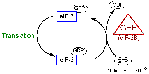
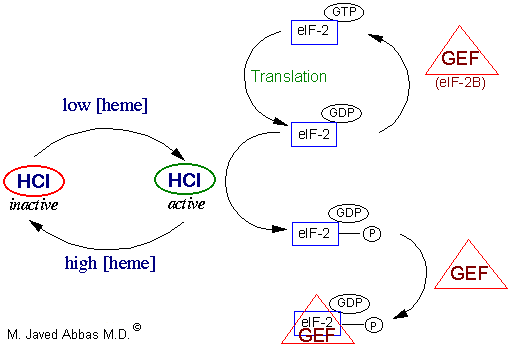
eIF-2B
(also called GEF)
GTP/GDP exchange during eIF-2 recycling
eIF-3
composed of ~10 subunits
ribosome subunit antiassociation, binding to 40S subunit
eIF-4F
composed of 3 subunits: eIF-4E, eIF-4A, eIF-4G
mRNA binding to 40S subunit, ATPase-dependent RNA helicase activity
eIF-4AATPase-dependent RNA helicase
eIF-4E5' cap recognition
eIF-4Gacts as a scaffold for the assembly of eIF-4E and -4A in the eIF-4F complex
eIF-4Bstimulates helicase, binds simultaneously with eIF-4F
eIF-5release of eIF-2 and eIF-3, ribosome-dependent GTPase
eIF-6ribosome subunit antiassociation

back to the top


Specific Steps in Translational Initiation
Initiation of translation requires 4 specific steps:
  • 1. A ribosome must dissociate into its' 40S and 60S subunits.
  • 2. A ternary complex termed the preinitiation complex is formed consisting of the initiator, GTP, eIF-2 and the 40S subunit.
  • 3. The mRNA is bound to the preinitiation complex.
  • 4. The 60S subunit associates with the preinitiation complex to form the 80S initiation complex.
The initiation factors eIF-1 and eIF-3 bind to the 40S ribosomal subunit favoring antiassociation to the 60S subunit. The prevention of subunit reassociation allows the preinitiation complex to form.
The first step in the formation of the preinitiation complex is the binding of GTP to eIF-2 to form a binary complex. eIF-2 is composed of three subunits, a, b and g. The binary complex then binds to the activated initiator tRNA, met-tRNAmet forming a ternary complex that then binds to the 40S subunit forming the 43S preinitiation complex. The preinitiation complex is stabilized by the earlier association of eIF-3 and eIF-1 to the 40S subunit.
The cap structure of eukaryotic mRNAs is bound by specific eIFs prior to association with the preinitiation complex. Cap binding is accomplished by the initiation factor eIF-4F. This factor is actually a complex of 3 proteins; eIF-4E, A and G. The protein eIF-4E is a 24 kDa protein which physically recognizes and binds to the cap structure. eIF-4A is a 46 kDa protein which binds and hydrolyzes ATP and exhibits RNA helicase activity. Unwinding of mRNA secondary structure is necessary to allow access of the ribosomal subunits. eIF-4G aids in binding of the mRNA to the 43S preinitiation complex.
Once the mRNA is properly aligned onto the preinitiation complex and the initiator met-tRNAmet is bound to the initiator AUG codon (a process facilitated by eIF-1) the 60S subunit associates with the complex. The association of the 60S subunit requires the activity of eIF-5 which has first bound to the preinitiation complex. The energy needed to stimulate the formation of the 80S initiation complex comes from the hydrolysis of the GTP bound to eIF-2. The GDP bound form of eIF-2 then binds to eIF-2B which stimulates the exchange of GTP for GDP on eIF-2. When GTP is exchanged eIF-2B dissociates from eIF-2. This is termed the eIF-2 cycle (see diagram below). This cycle is absolutely required in order for eukaryotic translational initiation to occur. The GTP exchange reaction can be affected by phosphorylation of the a-subunit of eIF-2.
At this stage the initiator met-tRNAmet is bound to the mRNA within a site of the ribosome termed the P-site, for peptide site. The other site within the ribosome to which incoming charged tRNAs bind is termed the A-site, for amino acid site.
The eIF-2 cycle involves the regeneration of GTP-bound eIF-2 following the hydrolysis of GTP during translational initiation. When the 40S preinitiation complex is engaged with the 60S ribosome to form the 80S initiation complex, the GTP bound to eIF-2 is hydrolyzed providing energy for the process. In order for additional rounds of translational initiation to occur, the GDP bound to eIF-2 must be exchanged for GTP. This is the function of eIF-2B which is also called guanine nucleotide exchange factor (GEF).

back to the top
Elongation
The process of elongation, like that of initiation requires specific non-ribosomal proteins. In E. coli these are EFs and in eukaryotes eEFs. Elongation of polypeptides occurs in a cyclic manner such that at the end of one complete round of amino acid addition the A site will be empty and ready to accept the incoming aminoacyl-tRNA dictated by the next codon of the mRNA. This means that not only does the incoming amino acid need to be attached to the peptide chain but the ribosome must move down the mRNA to the next codon. Each incoming aminoacyl-tRNA is brought to the ribosome by an eEF-1a-GTP complex. When the correct tRNA is deposited into the A site the GTP is hydrolyzed and the eEF-1a-GDP complex dissociates. In order for additional translocation events the GDP must be exchanged for GTP. This is carried out by eEF-1bg similarly to the GTP exchange that occurs with eIF-2 catalyzed by eIF-2B.
The peptide attached to the tRNA in the P site is transferred to the amino group at the aminoacyl-tRNA in the A site. This reaction is catalyzed by peptidyltransferase. This process is termed transpeptidation. The elongated peptide now resides on a tRNA in the A site. The A site needs to be freed in order to accept the next aminoacyl-tRNA. The process of moving the peptidyl-tRNA from the A site to the P site is termed, translocation. Translocation is catalyzed by eEF-2 coupled to GTP hydrolysis. In the process of translocation the ribosome is moved along the mRNA such that the next codon of the mRNA resides under the A site. Following translocation eEF-2 is released from the ribosome. The cycle can now begin again.
back to the top
Termination
Like initiation and elongation, translational termination requires specific protein factors identified as releasing factors, RFs in E. coli and eRFs in eukaryotes. There are 2 RFs in E. coli and one in eukaryotes. The signals for termination are the same in both prokaryotes and eukaryotes. These signals are termination codons present in the mRNA. There are 3 termination codons, UAG, UAA and UGA.
In E. coli the termination codons UAA and UAG are recognized by RF-1, whereas RF-2 recognizes the termination codons UAA and UGA. The eRF binds to the A site of the ribosome in conjunction with GTP. The binding of eRF to the ribosome stimulates the peptidytransferase activity to transfer the peptidyl group to water instead of an aminoacyl-tRNA. The resulting uncharged tRNA left in the P site is expelled with concomitant hydrolysis of GTP. The inactive ribosome then releases its mRNA and the 80S complex dissociates into the 40S and 60S subunits ready for another round of translation.
back to the top
Selenoproteins
Selenium is a trace element and is found as a component of several prokaryotic and eukaryotic enzymes that are involved in redox reactions. The selenium in these selenoproteins is incorporated as a unique amino acid, selenocysteine, during translation. A particularly important eukaryotic selenoenzyme is glutathione peroxidase. This enzyme is required during the oxidation of glutathione by hydrogen peroxide (H2O2) and organic hydroperoxides.
Structure of the selenocysteine residue
Incorporation of selenocysteine by the translational machinery occurs via an interesting and unique mechanism. The tRNA for selenocysteine is charged with serine and then enzymatically selenylated to produce the selenocysteinyl-tRNA. The anticodon of selenocysteinyl-tRNA interacts with a stop codon in the mRNA (UGA) instead of a serine codon. The selenocysteinyl-tRNA has a unique structure that is not recognized by the termination machinery and is brought into the ribosome by a dedicated specific elongation factor. An element in the 3' non-translated region (UTR) of selenoprotein mRNAs determines whether UGA is read as a stop codon or as a selenocysteine codon.
back to the top
Heme Control of Translation
Regulation of initiation in eukaryotes is effected by phosphorylation of a ser(S) residue in the a subunit of eIF-2. Phosphorylated eIF-2 in the absence of eIF-2B is just as active an initiator as non-phosphorylated eIF-2. However, when eIF-2 is phosphorylated the GDP-bound complex is stabilized and exchange for GTP is inhibited. The exchange of GDP for GTP is mediated by eIF-2B (also called guanine nucleotide exchange factor - GEF). When eIF-2 is phosphorylated it binds eIF-2B more tightly thus slowing the rate of exchange. It is this inhibited exchange that affects the rate of initiation.
The phosphorylation of eIF-2 is the result of an activity called heme-controlled inhibitor (HCI) which functions as diagrammed below. HCI is generated in the absence of heme, a mitochondrial product. Removal of phosphate is catalyzed by a specific eIF-2 phosphatase which is unaffected by heme. The presence of HCI was first seen in in vitro translation system derived from lysates of reticulocytes. Reticulocytes synthesize almost exclusively hemoglobin at an extremely high rate. In an intact reticulocyte eIF-2 is protected from phosphorylation by a specific 67 kDa protein.
The regulation of translation by heme controlled inhibitor (HCI). Control of translation by heme is clinically important only in erythrocytes. Erythrocytes are enucleate and contain primarily globin mRNA. When the level of heme (required for the synthesis of biologically active hemoglobin) is low it would be inefficient for erythrocytes to synthesize globin protein. As the level of heme falls the activity of HCI increases. HCI is a kinase which phosphorylates eIF-2. When phosphorylated, eIF-2 still hydrolyzes bound GTP to GDP and still interacts with eIF-2B (GEF). However, the rate of eIF-2B-mediated GTP exchange is greatly reduced. This renders eIF-2 incapable of being used to form an new ternary initiation complex and translational initiation is reduced. When the level of heme again rises the activity of HCI is reduced and translational initiation is once again active.

back to the top
Interferon Control of Translation
Regulation of translation can also be induced in virally infected cells. It would benefit a virally infected cell to turn off protein synthesis to prevent propagation of the viruses. This is accomplished by the induced synthesis of interferons (IFs). There are 3 classes of IFs. The leukocyte or a-IFs, the fibroblast or b-IFs and the lymphocyte or g-IFs. IFs are induced by dsRNAs and themselves induce a specific kinase termed RNA-dependent protein kinase (PKR) that phosphorylates eIF-2 thereby shutting off translation in a similar manner to that of heme control of translation. Additionally, IFs induce the synthesis of 2'-5'-oligoadenylate, pppA(2'p5'A)n, that activates a pre-existing ribonuclease, RNase L. RNase L degrades all classes of mRNAs thereby shutting off translation.
back to the top
Iron Control of Translation
Regulation of the translation of certain mRNAs occurs through the action of specific RNA-binding proteins. Protein of this class have been identified that bind to sequences in either the 5' non-translated region (5'-UTR) or 3'-UTR. Two particularly interesting and important regulatory schemes related to iron metabolism encompass RNA binding proteins that bind to either the 5'-UTR of one mRNA or the 3'-UTR of another.
The transferrin receptor is a protein located in the plasma membrane that binds the protein transferrin. Transferrin is the major iron transport protein in the plasma. When iron levels are low the rate of synthesis of the transferrin receptor mRNA increases so that cells can take up more iron. This regulation occurs through the action of an iron response element binding protein (IRBP) that binds to specific iron response elements (IREs) in the 3'-UTR of the transferrin receptor mRNA. These IREs form hair-pin loop structures that are recognized by IRBP. This IRBP is an iron-deficient form of aconitase, the iron-requiring enzyme of the TCA cycle. When iron levels are low, IRBP is free of iron and can therefore, interact with the IREs in the 3'-UTR of the transferrin receptor mRNA. Transferrin receptor mRNA with IRBP bound is stabilized from degradation. Conversely, when iron levels are high, IRBP binds iron then cannot interact with the IREs in the transferrin receptor mRNA. The effect is an increase in degradation of the transferrin receptor mRNA.
A related, but opposite, phenomenon controls the translation of the ferritin mRNA. Ferritin is an iron-binding protein that prevents toxic levels of ionized iron (Fe2+) from building up in cells. The ferritin mRNA has an IRE in its 5'-UTR. As with the transferrin receptor story, when iron levels are high, IRBP cannot bind to the IRE in the 5'-UTR of the ferritin mRNA. This allows the ferritin mRNA to be translated. Conversely, when iron levels are low, the IRBP binds to the IRE in the ferrritin mRNA preventing its translation.

back to the top
Protein Synthesis Inhibitors
Many of the antibiotics utilized for the treatment of bacterial infections as well as certain toxins function through the inhibition of translation. Inhibition can be effected at all stages of translation from initiation to elongation to termination.

Several Antibiotic and Toxin inhibitors of Translation

InhibitorComments
Chloramphenicolinhibits prokaryotic peptidyl transferase
Streptomycininhibits prokaryotic peptide chain initiation, also induces mRNA misreading
Tetracyclineinhibits prokaryotic aminoacyl-tRNA binding to the ribosome small subunit
Neomycinsimilar in activity to streptomycin
Erythromycininhibits prokaryotic translocation through the ribosome large subunit
Fusidic acidsimilar to erythromycin only by preventing EF-G from dissociating from the large subunit
Puromycinresembles an aminoacyl-tRNA, interferes with peptide transfer resulting in premature termination in both prokaryotes and eukaryotes
Diptheria toxincatalyzes ADP-ribosylation of and inactivation of eEF-2
Ricinfound in castor beans, catalyzes cleavage of the eukaryotic large subunit rRNA
Cycloheximideinhibits eukaryotic peptidyltransferase

back to the top
Back to Topics<<<<


This article has been modified by Dr. M. Javed Abbas.
If you have any comments please do not hesitate to sign my Guest Book.

20:50 21/12/2002